


Hi all
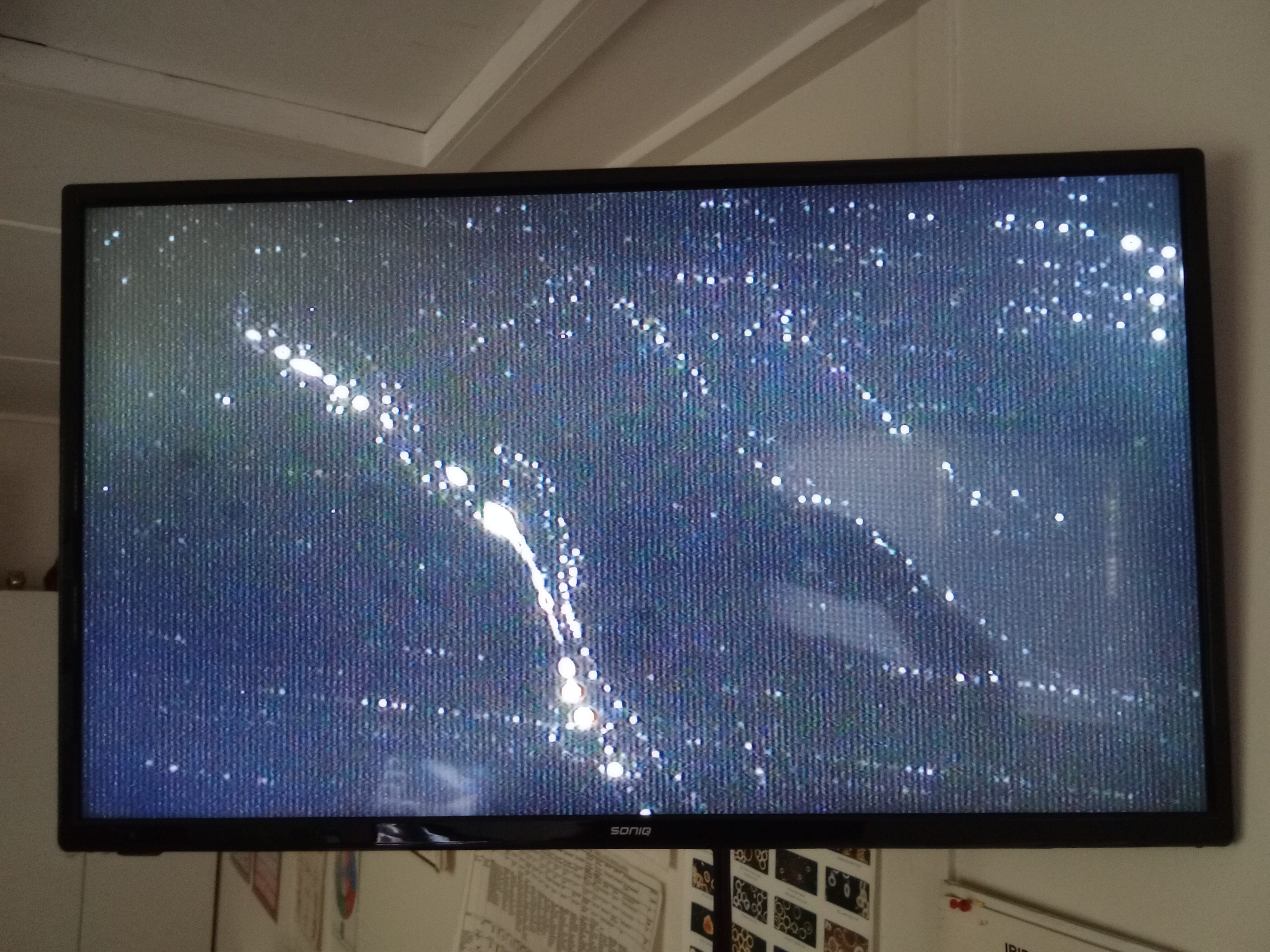
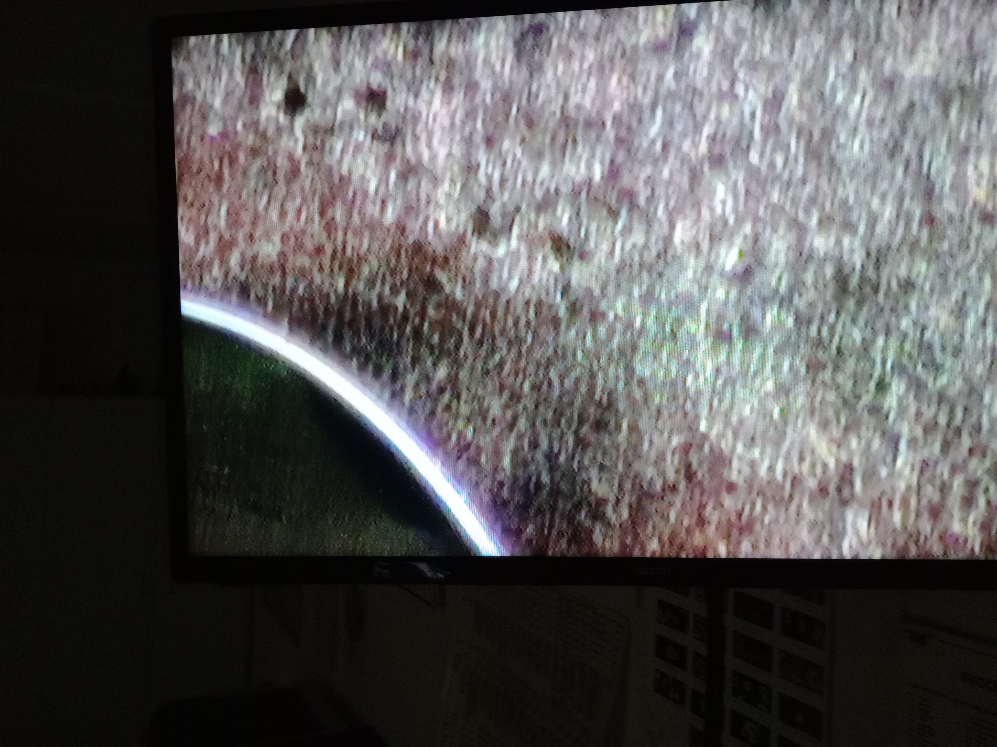
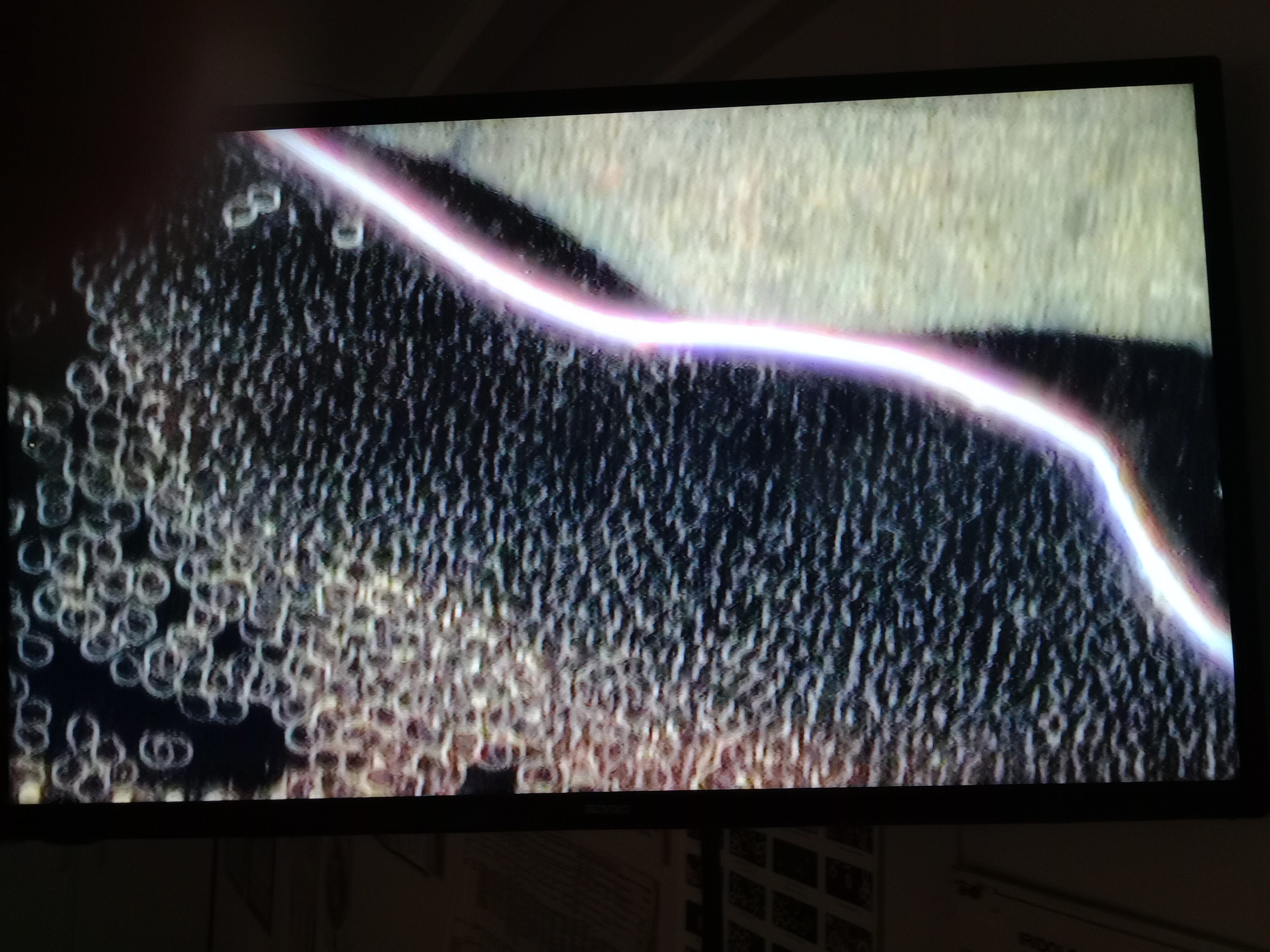
I try to get back every rnr and assess for change. Now and then I'll see a change that appears obvious such as the following lines.
As I said previously there us something to not getting as close up as you can because you may miss the big picture.
I've bought a spooky 2 and started with that as well as the skio work I do. On top of that I'm well wormed, reintroduced the glutathione, still use nac and colloidal gold.
Also use alot of Natto/kinese supplement and have now introduced dandelion leaf extract so going forward it will be interesting to see how much rouleaux and clotting I'll display.
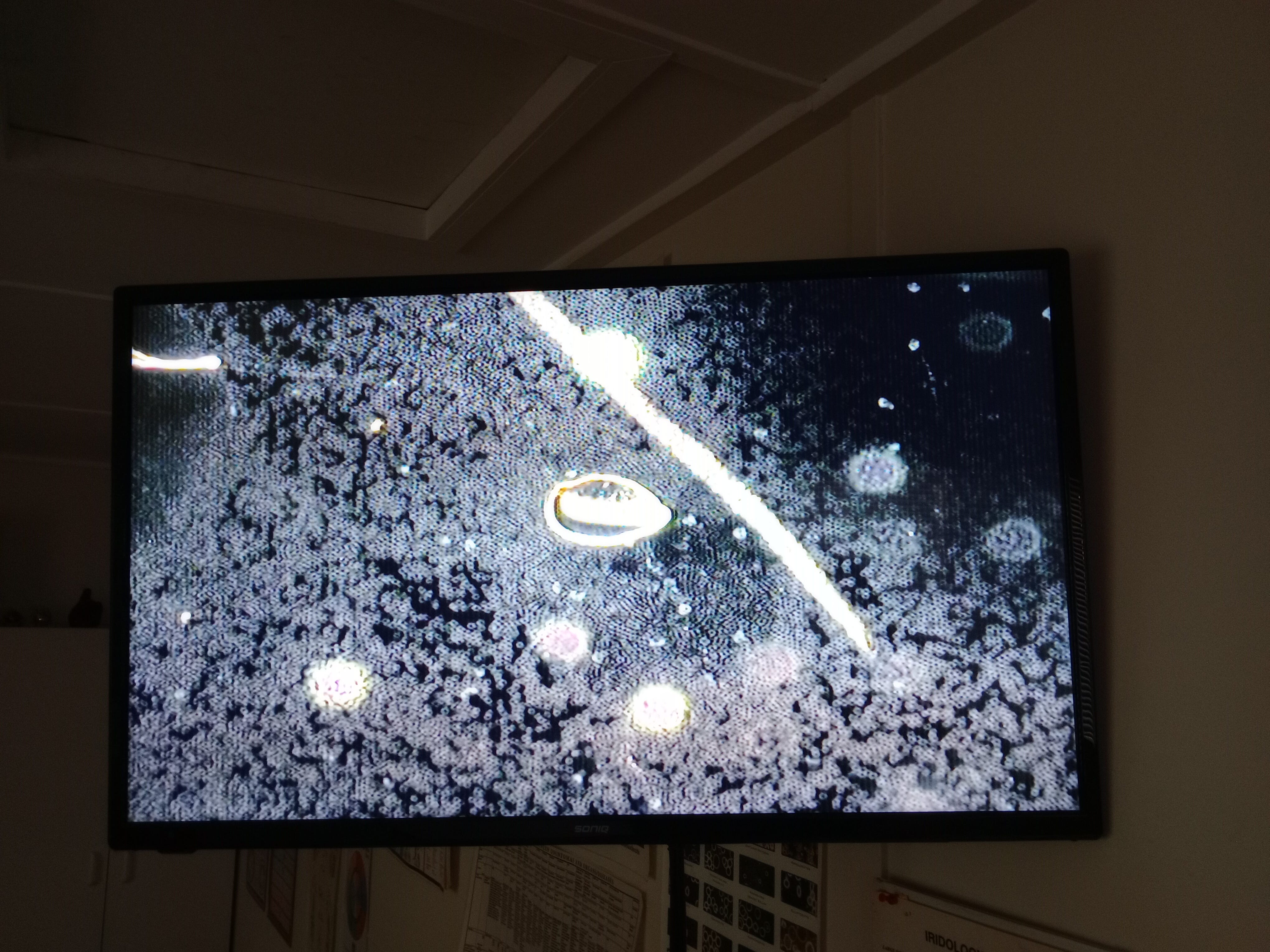
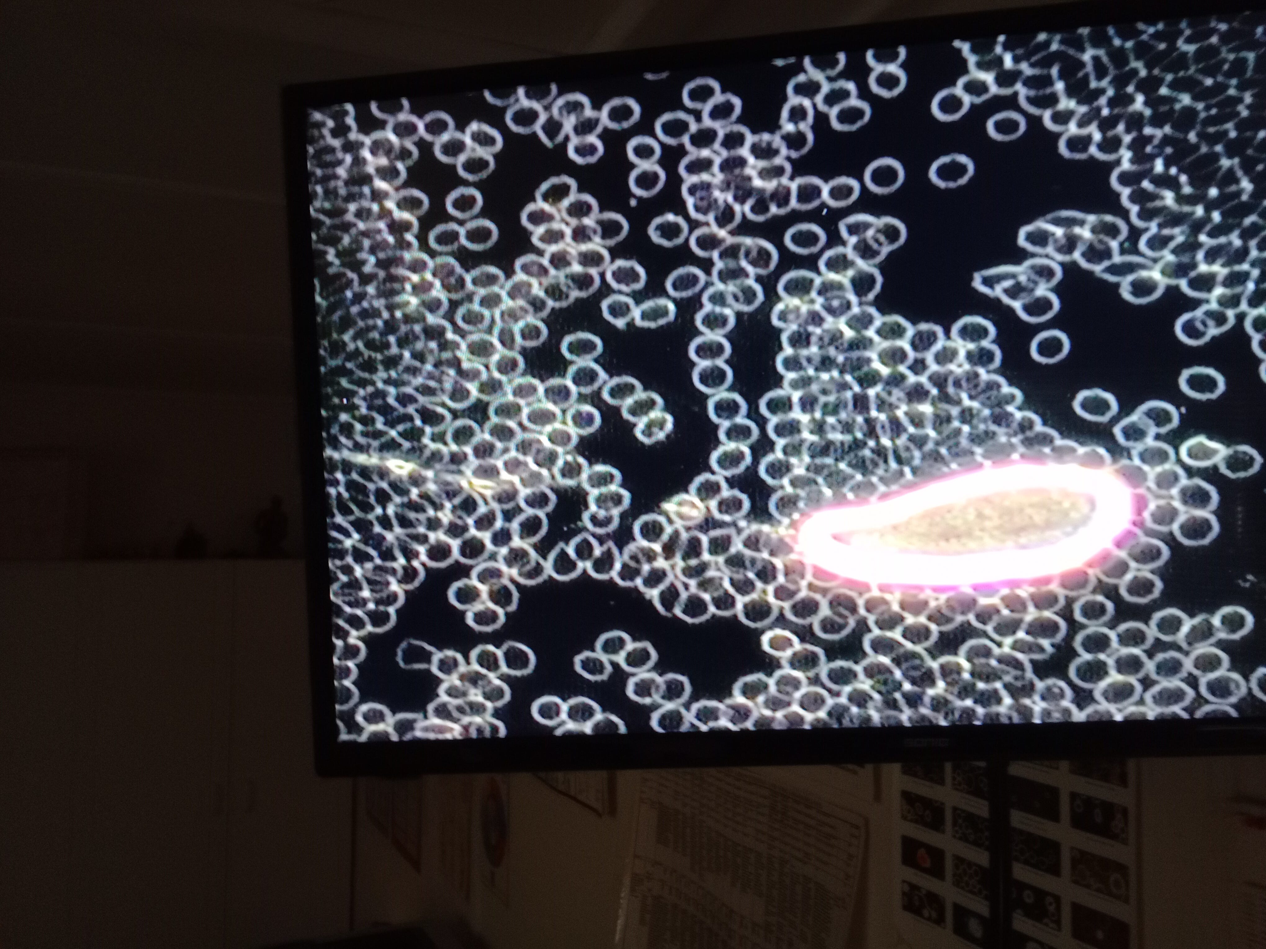
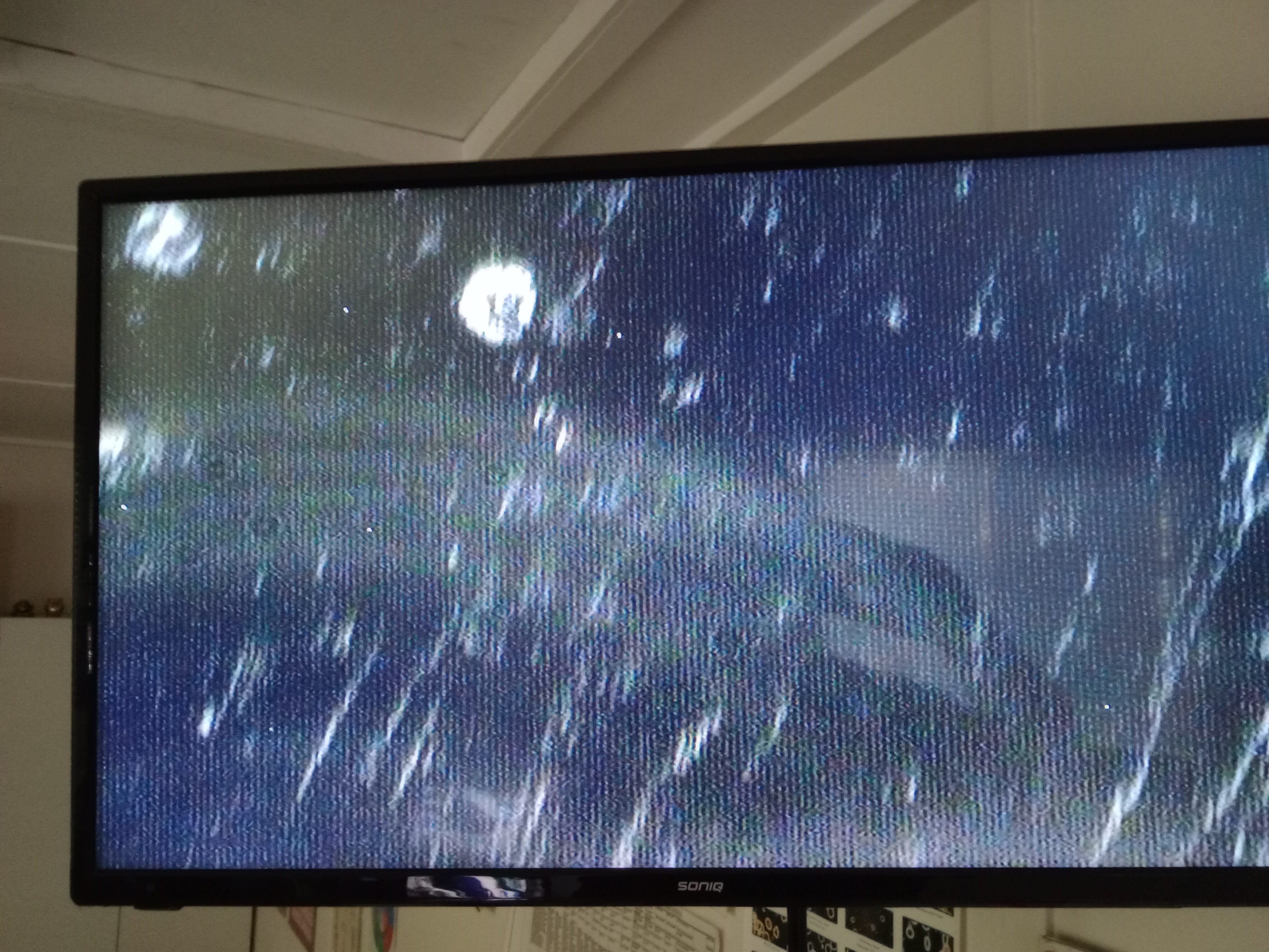
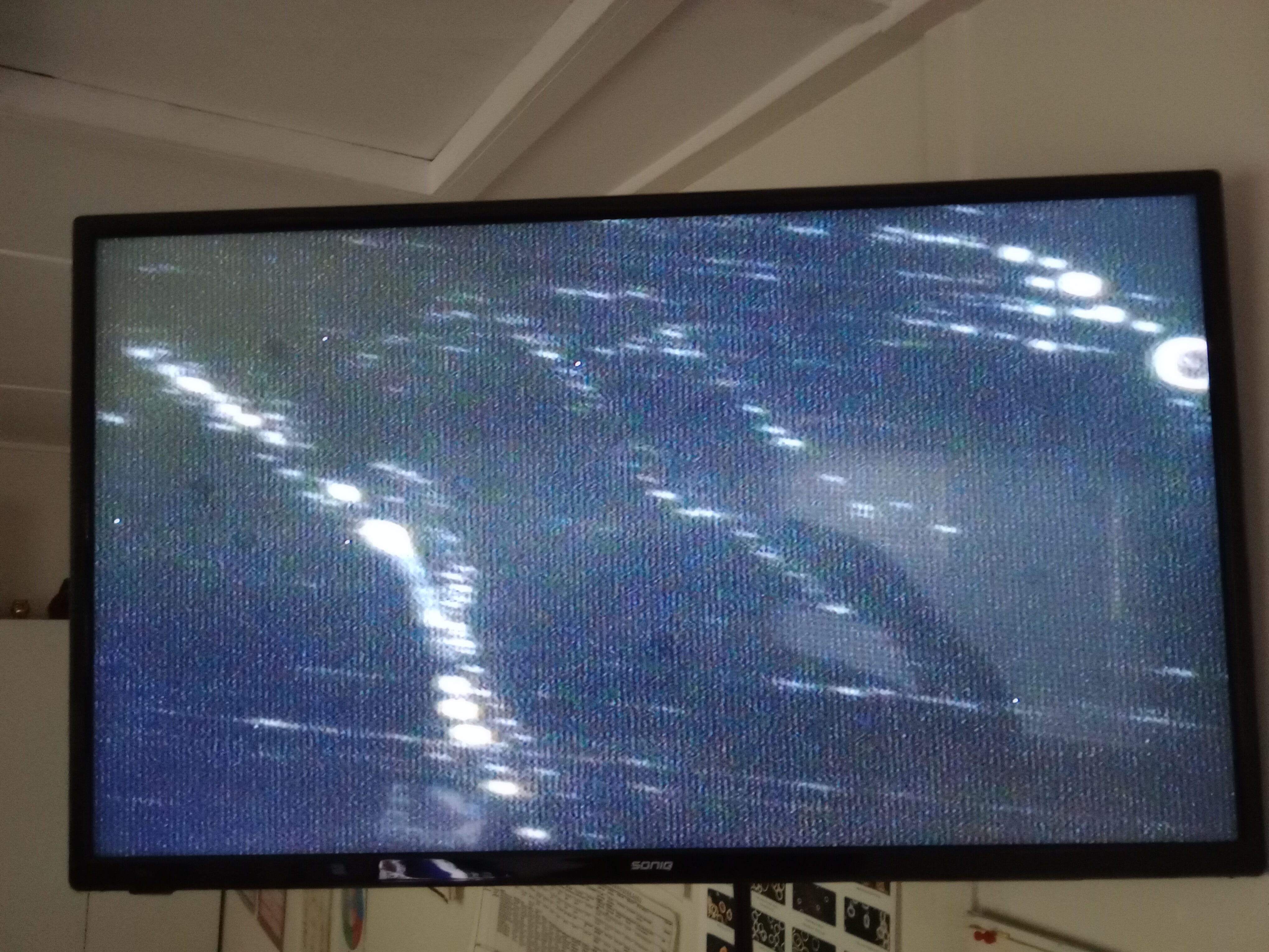
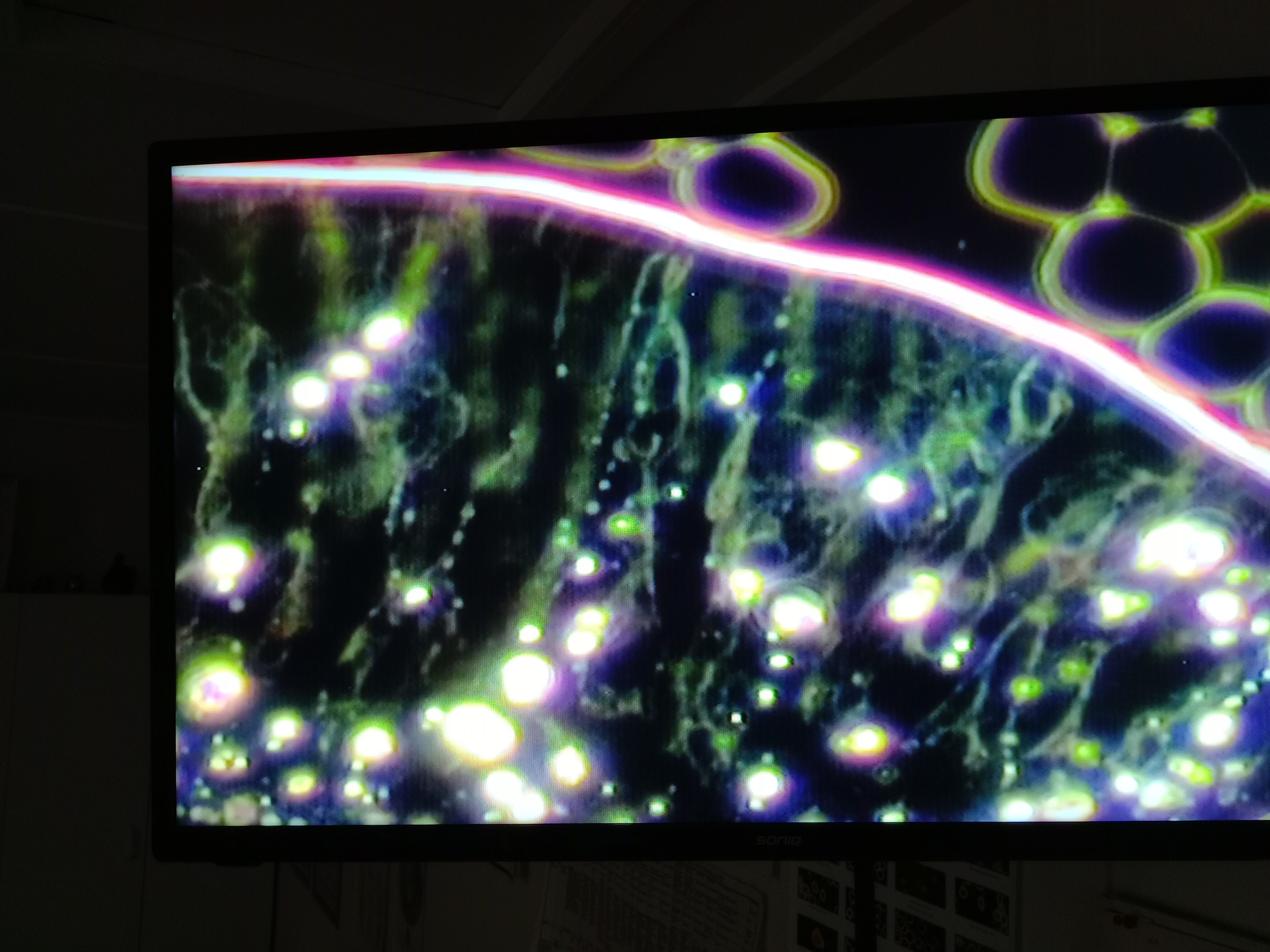
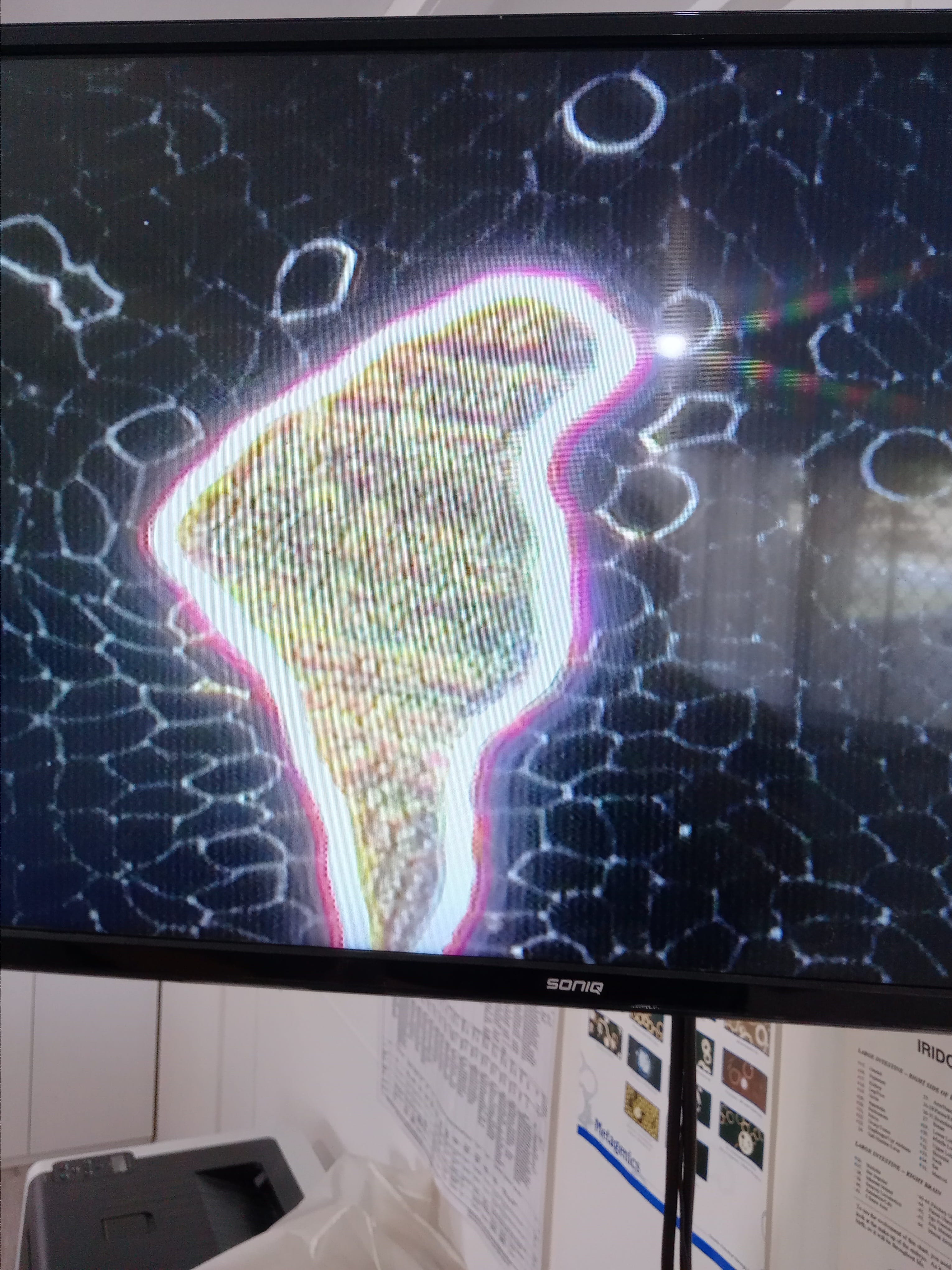
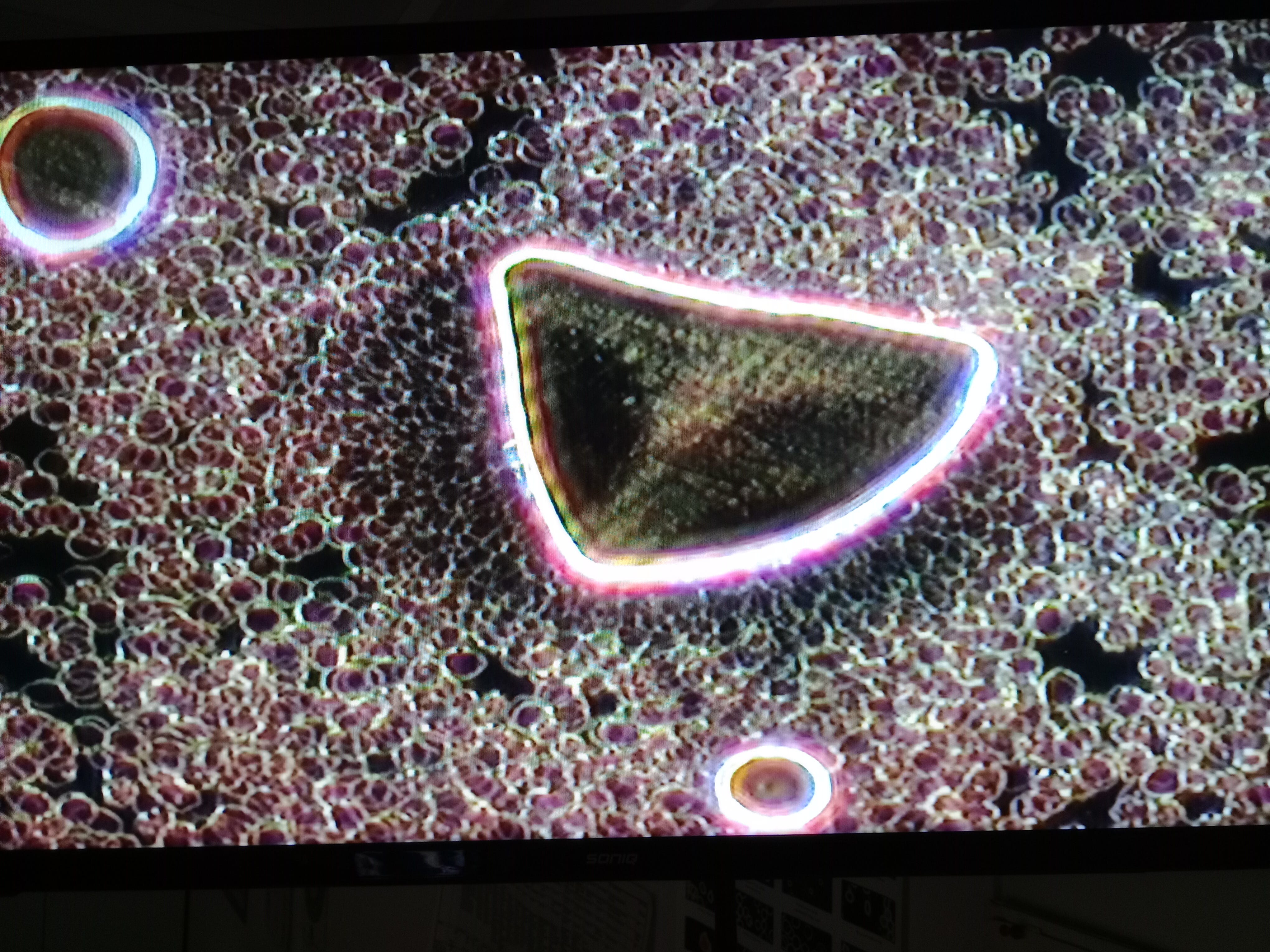
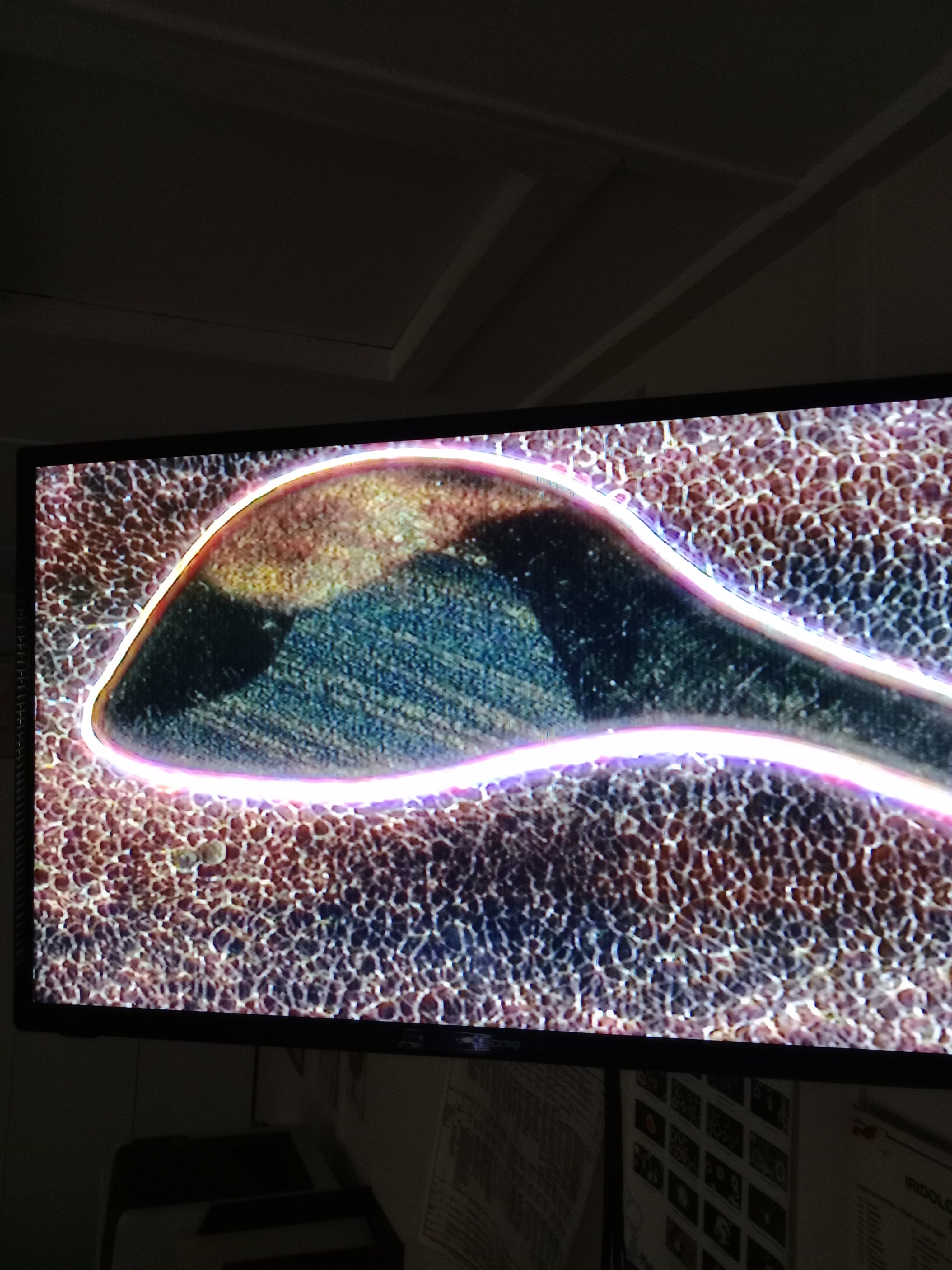
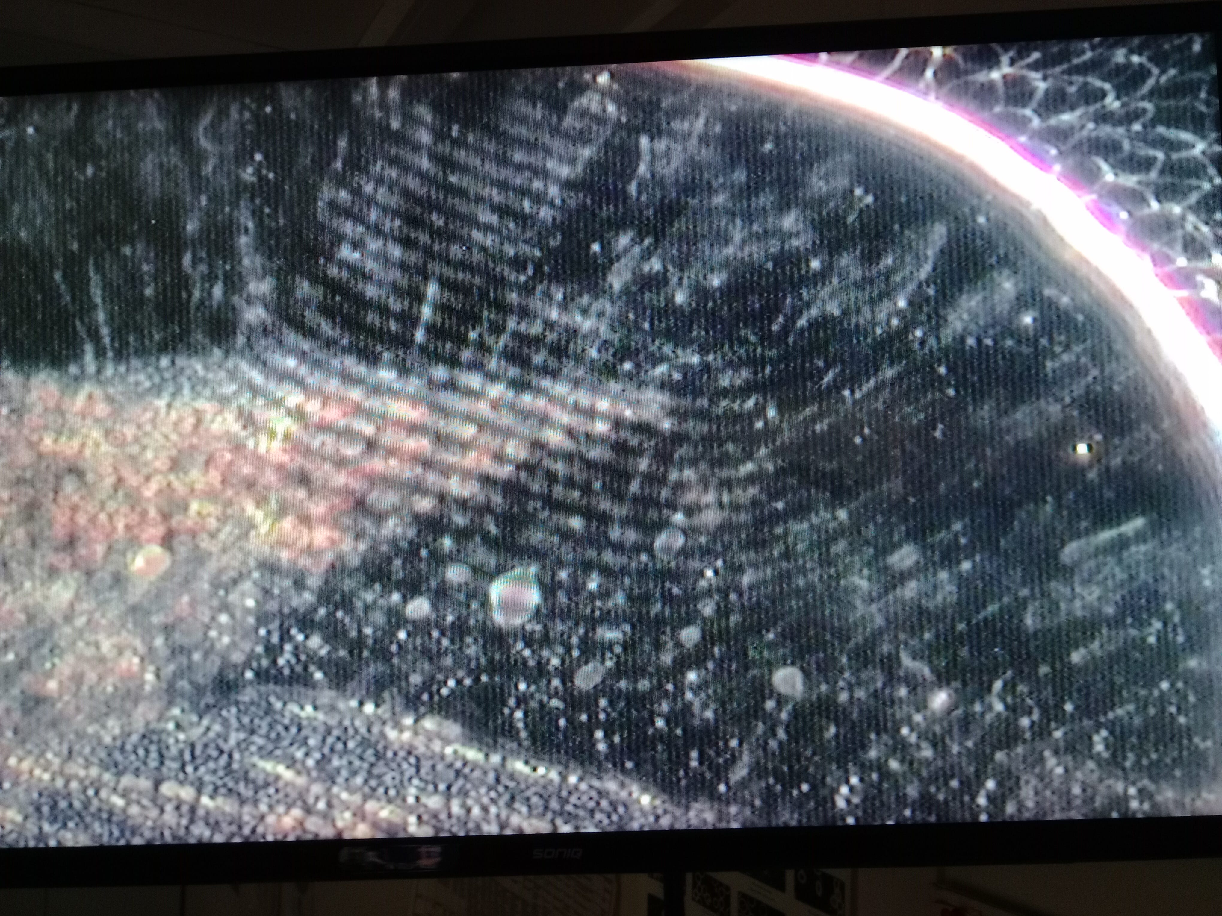
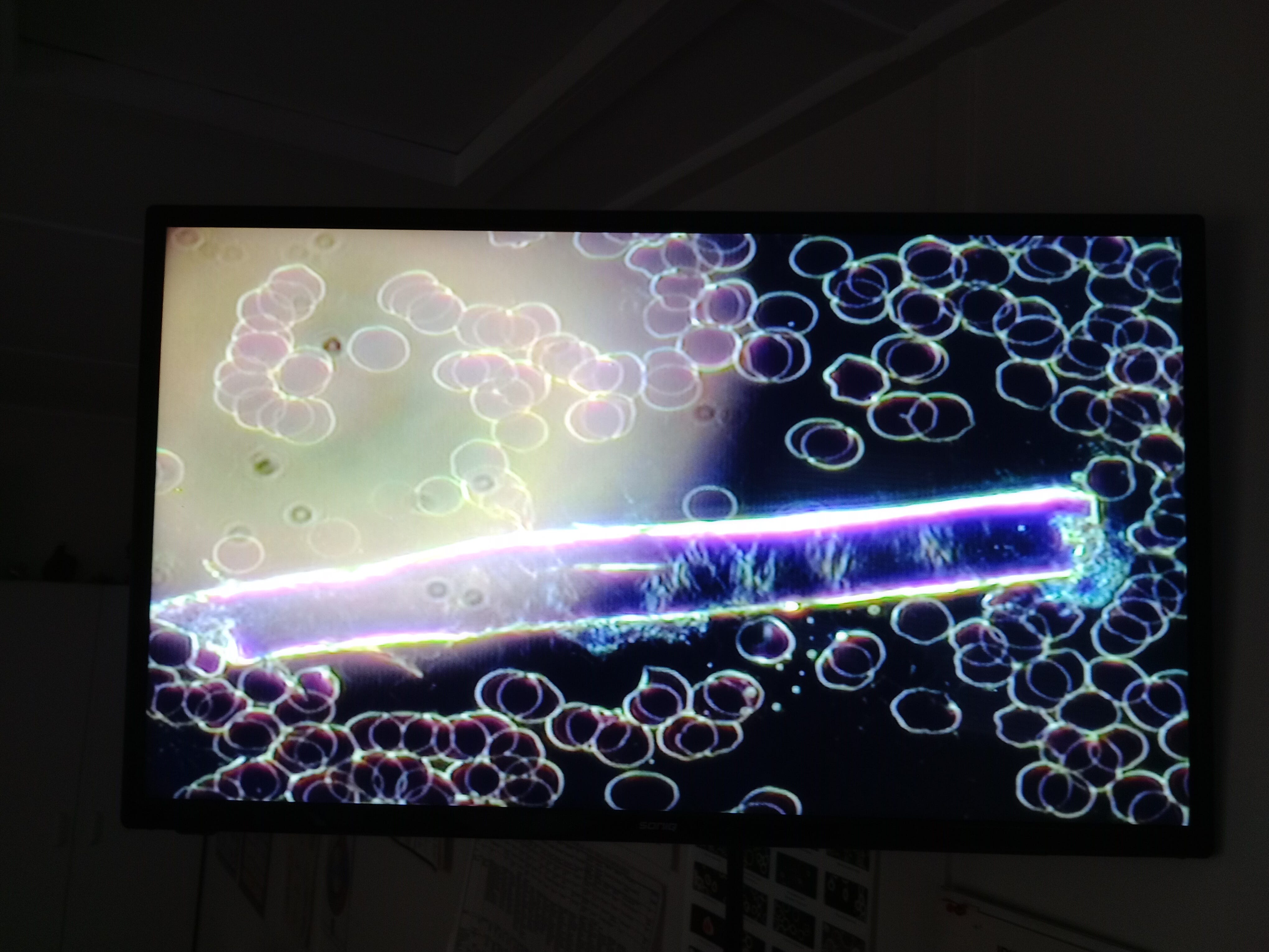
This is a close up of the first photo, all I have is speculation at the moment but if nothing else I've never seen it before or my blood analyst (and he’s been doing this for near on 30 years).
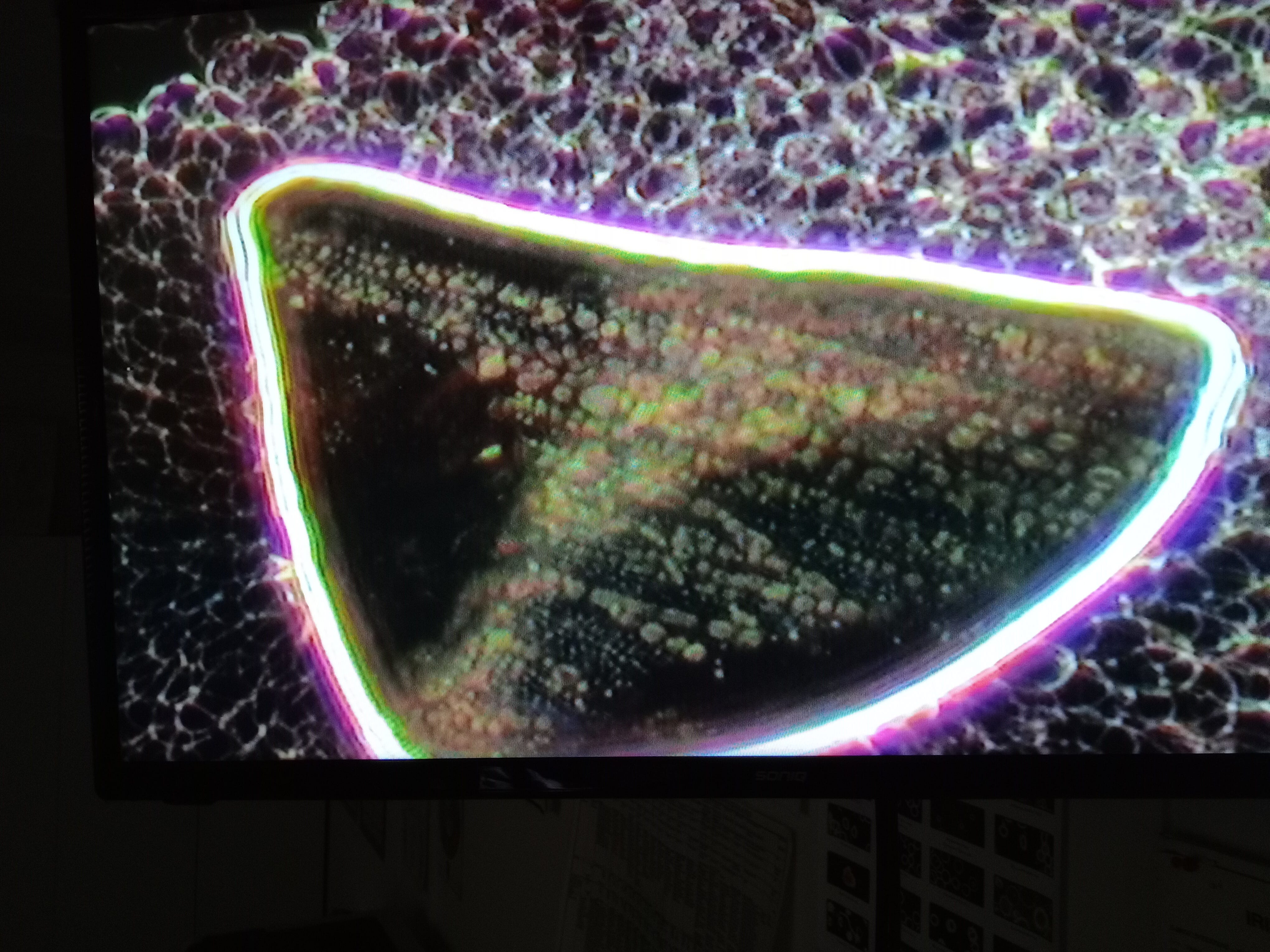
The thick line up close and as it tapers out.
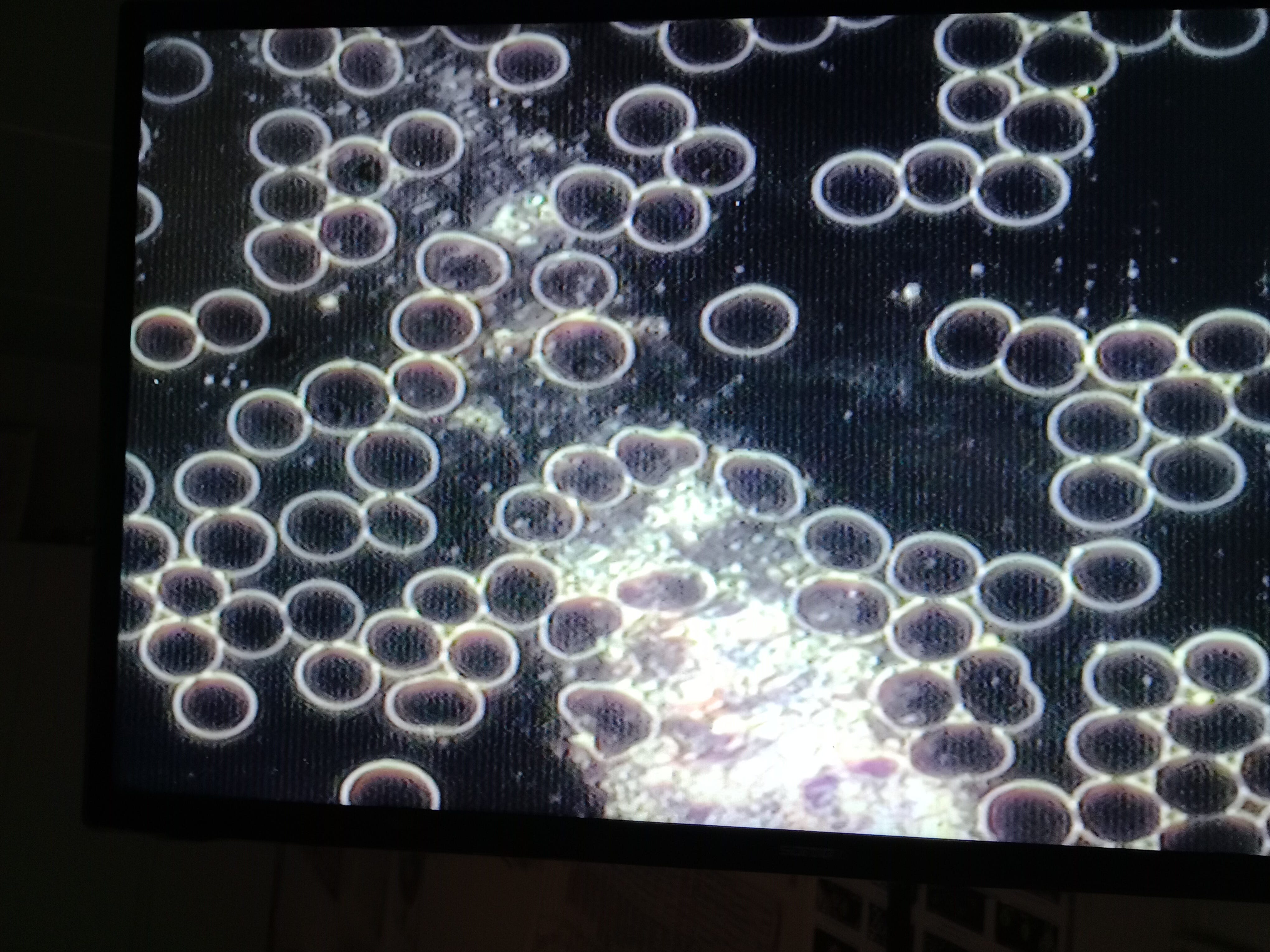
Thats the big think line (whatever it is) but also of note is the finer filigree threading through the rbc's. Looks like uts starting to link up the bubbles, ir at least links back to one.

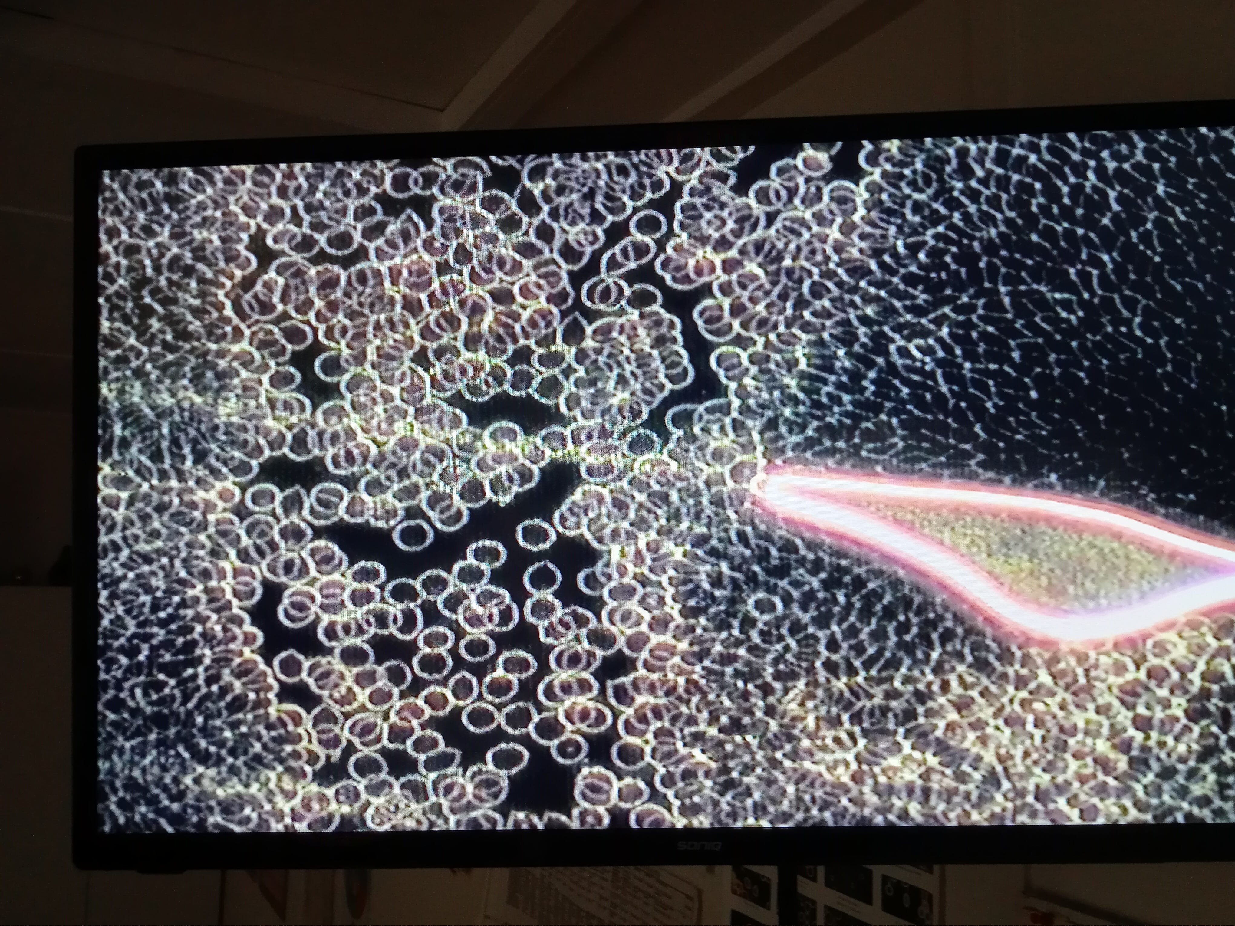
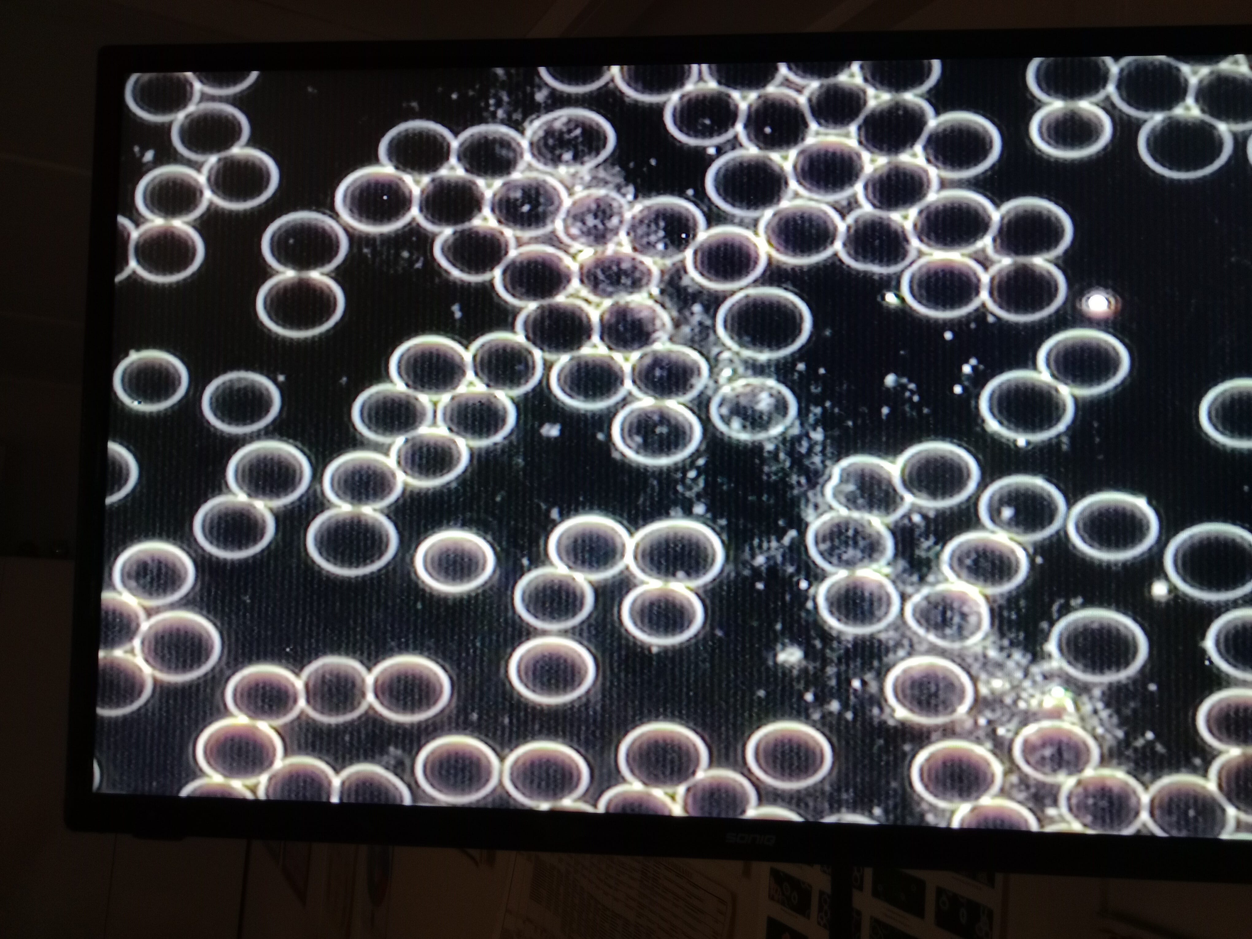
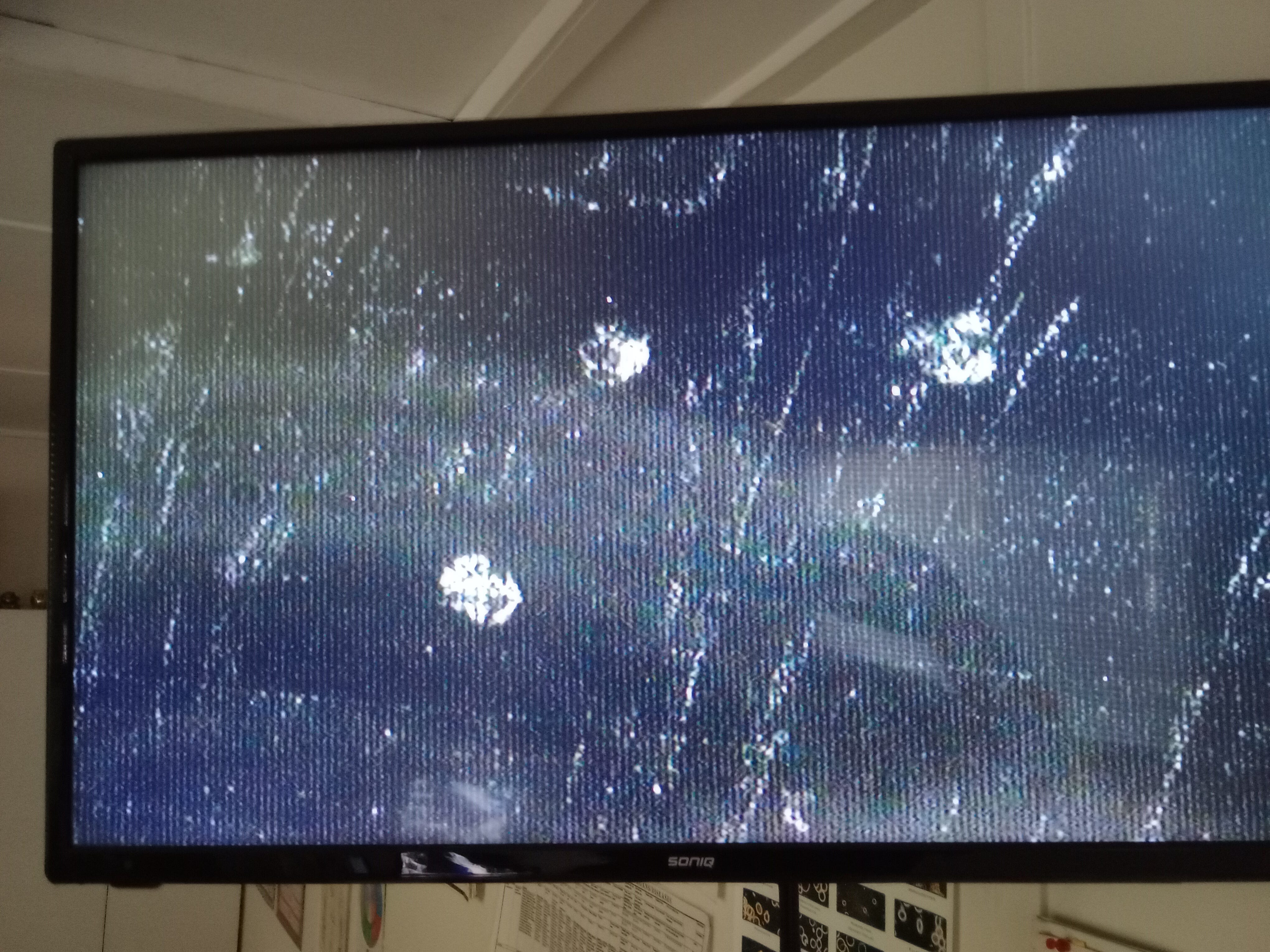
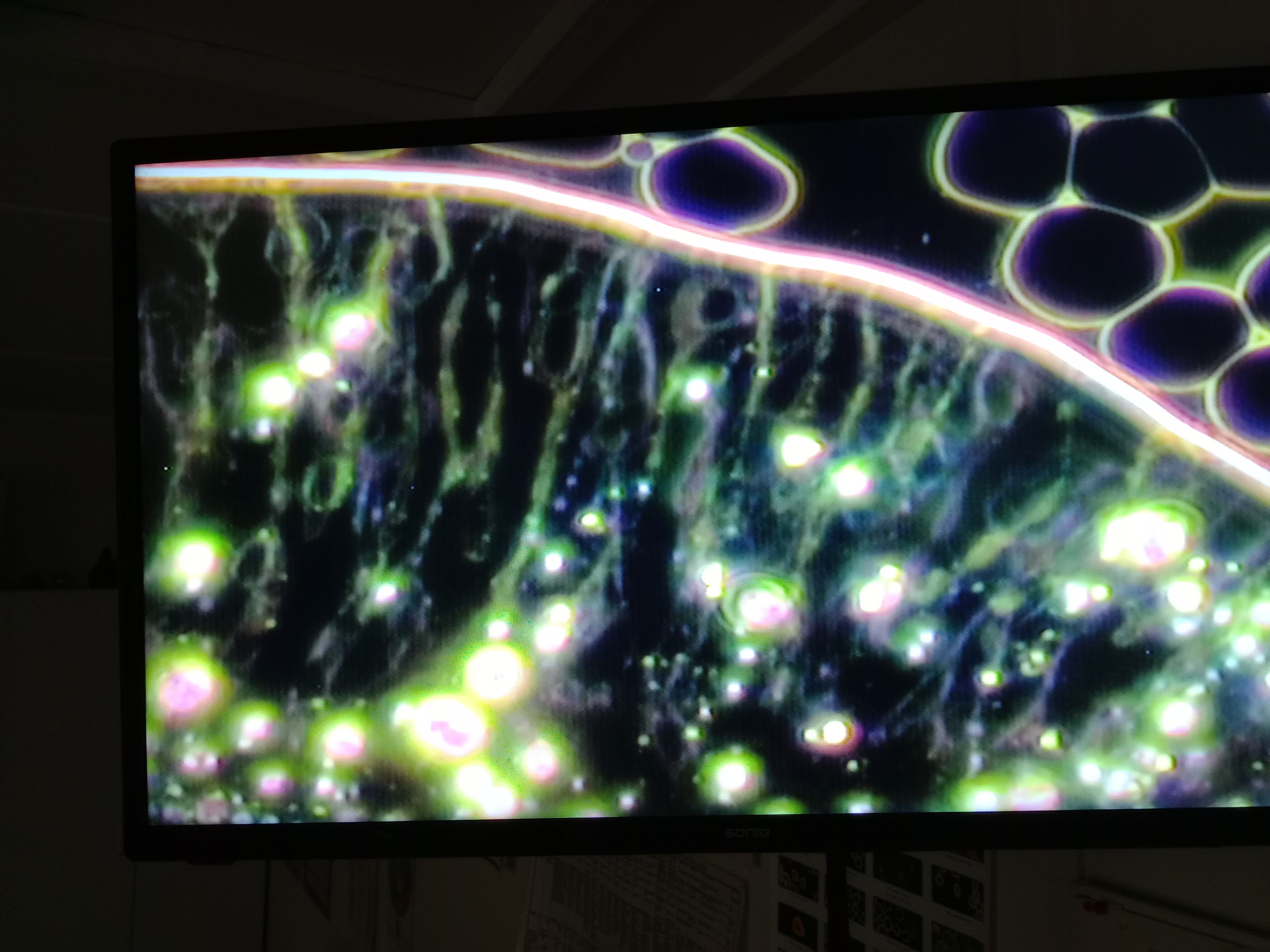
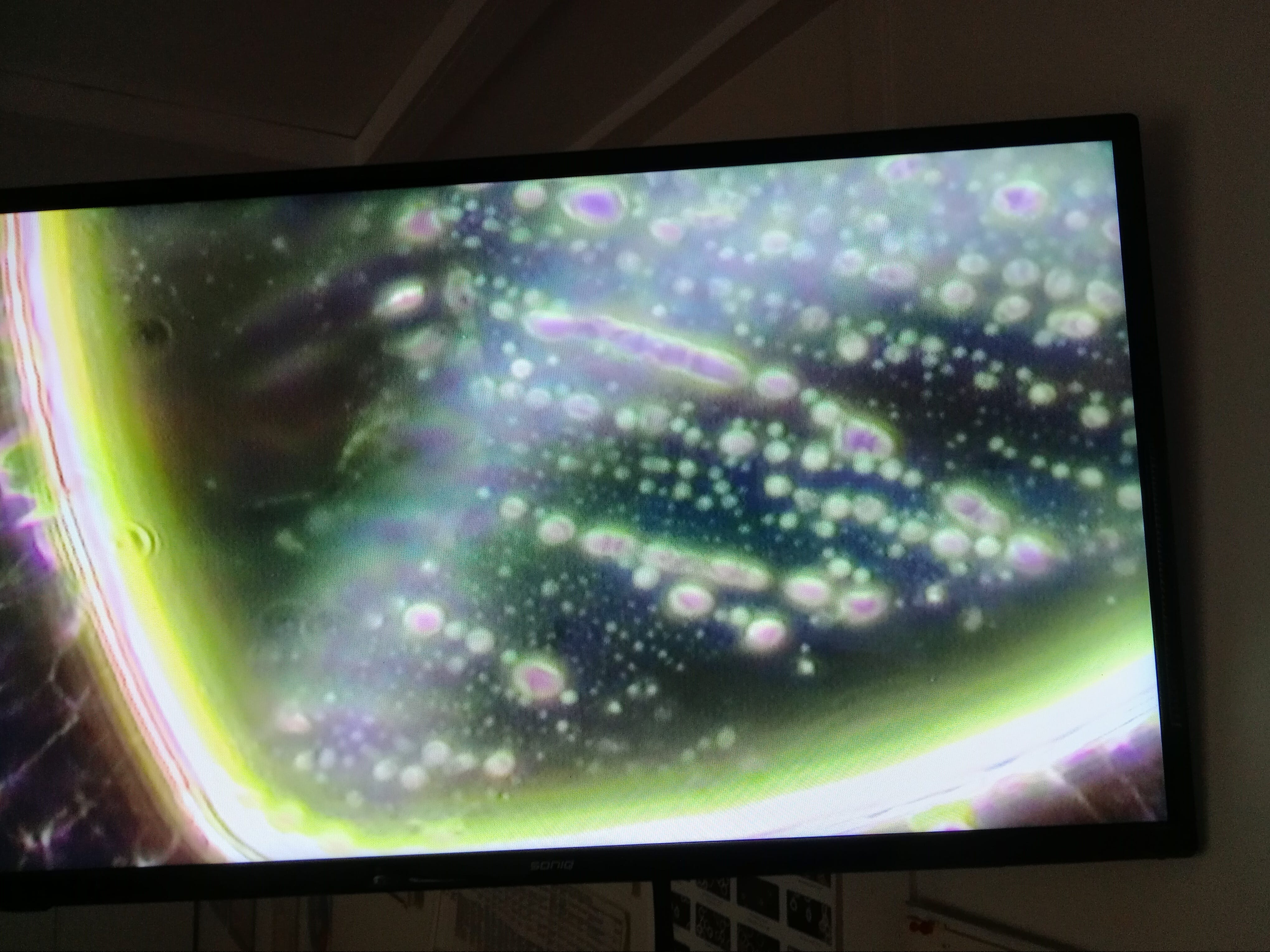
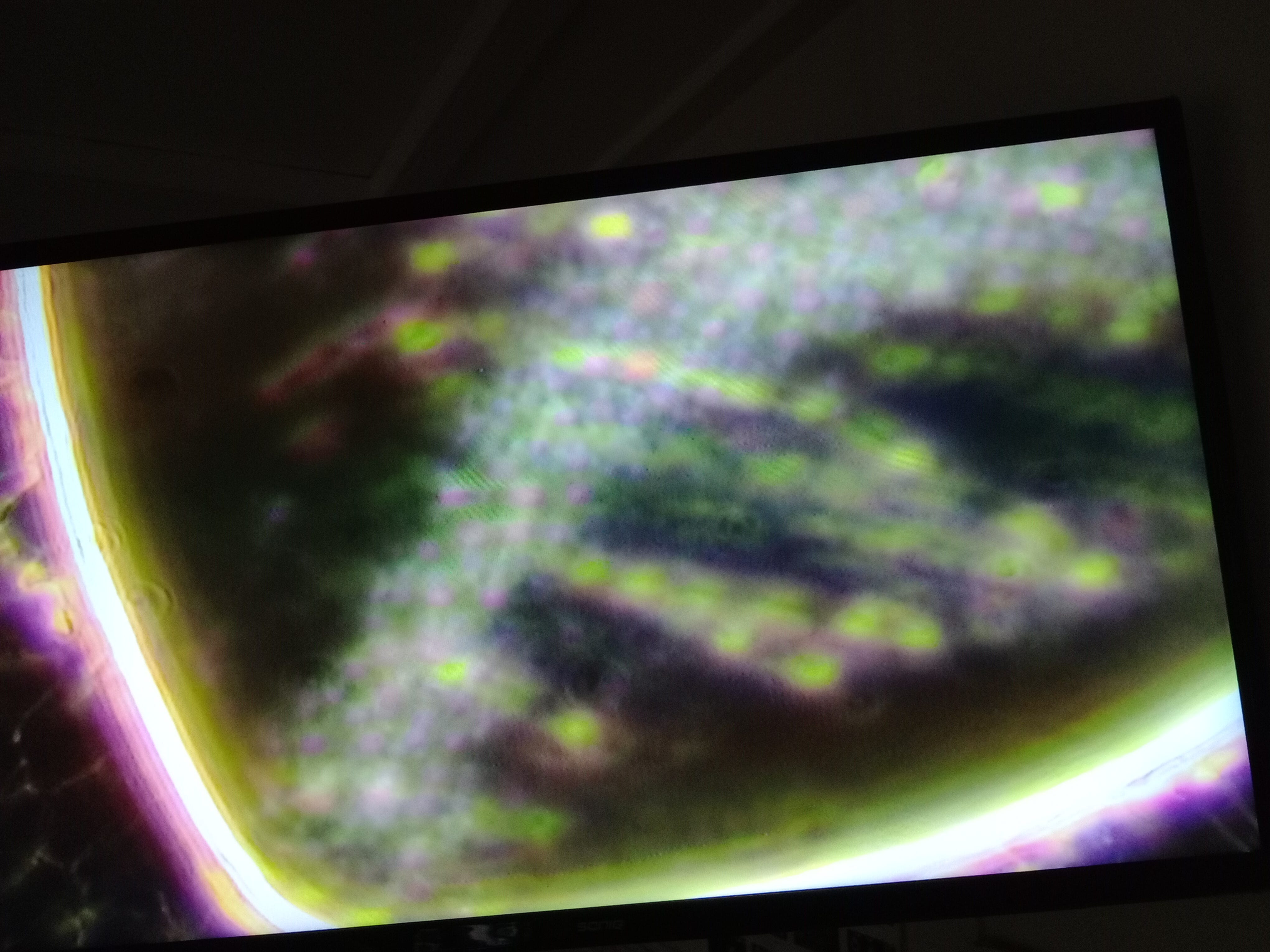
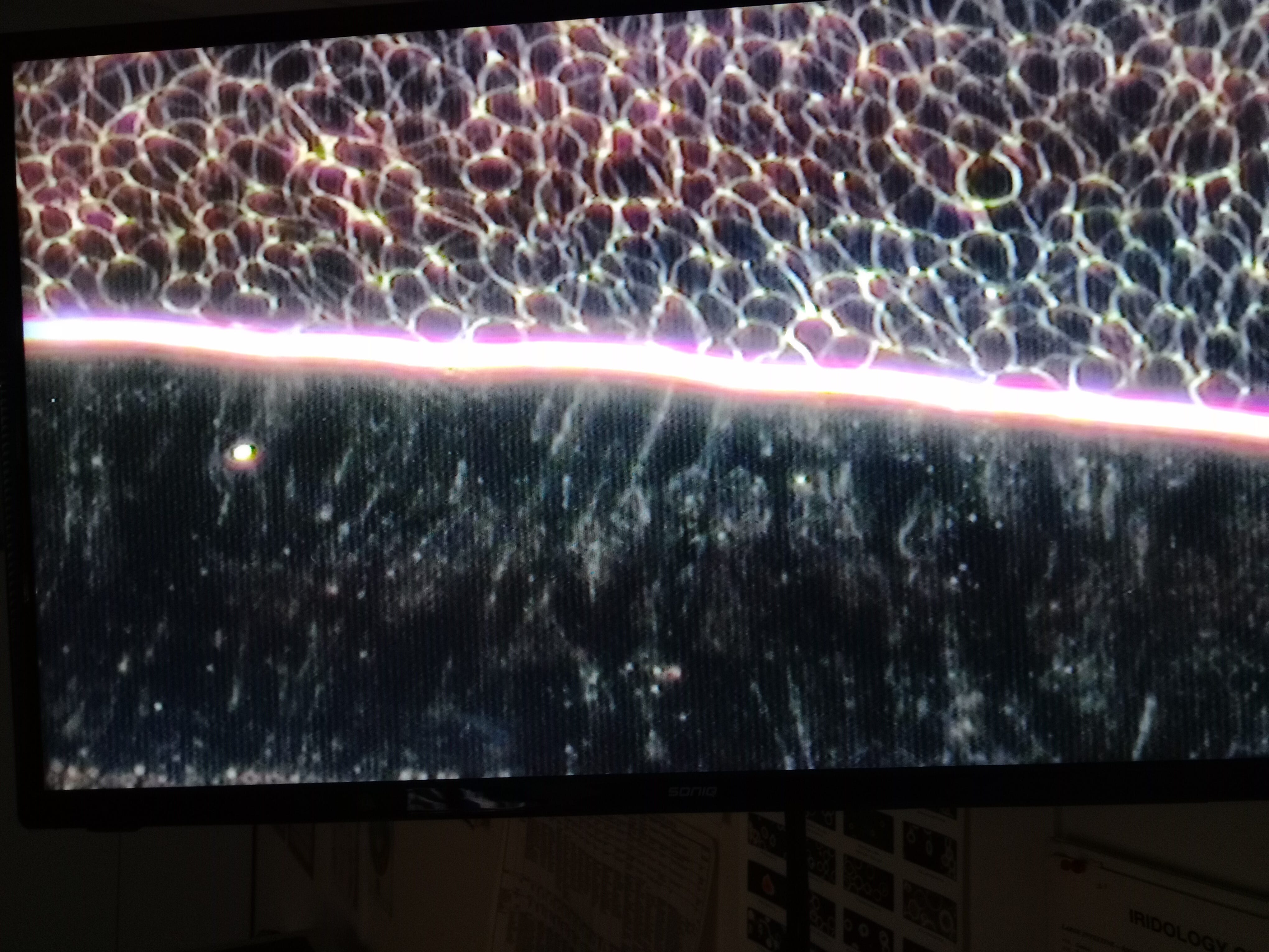
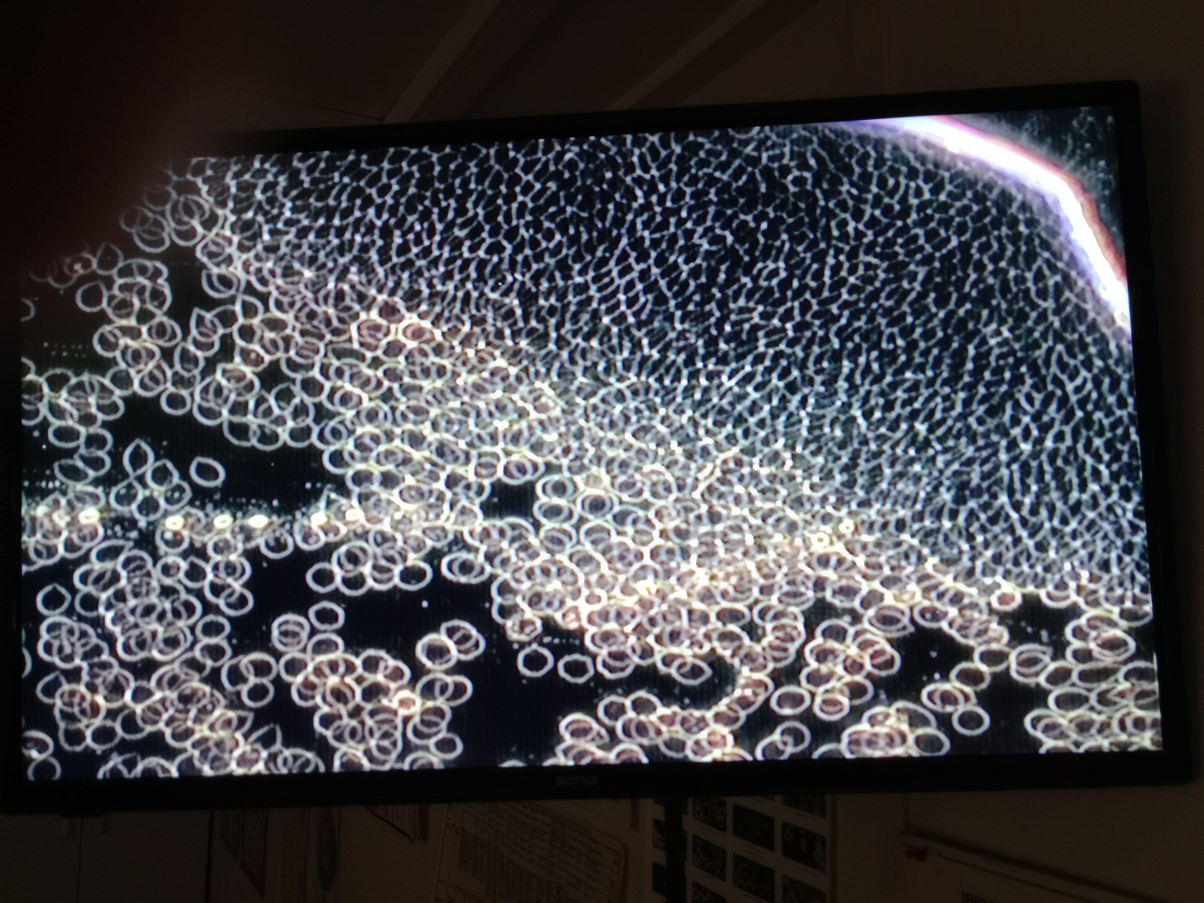
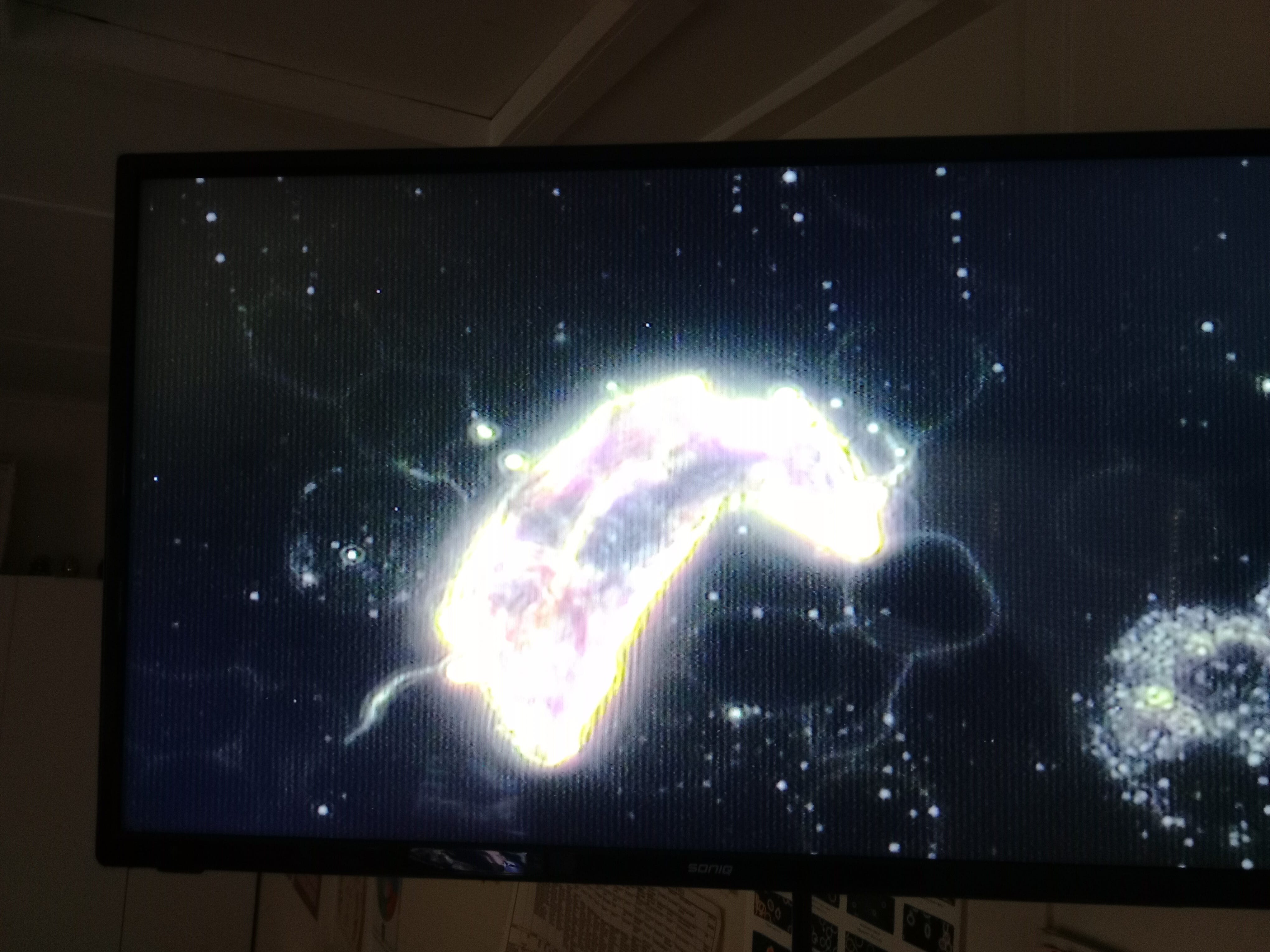
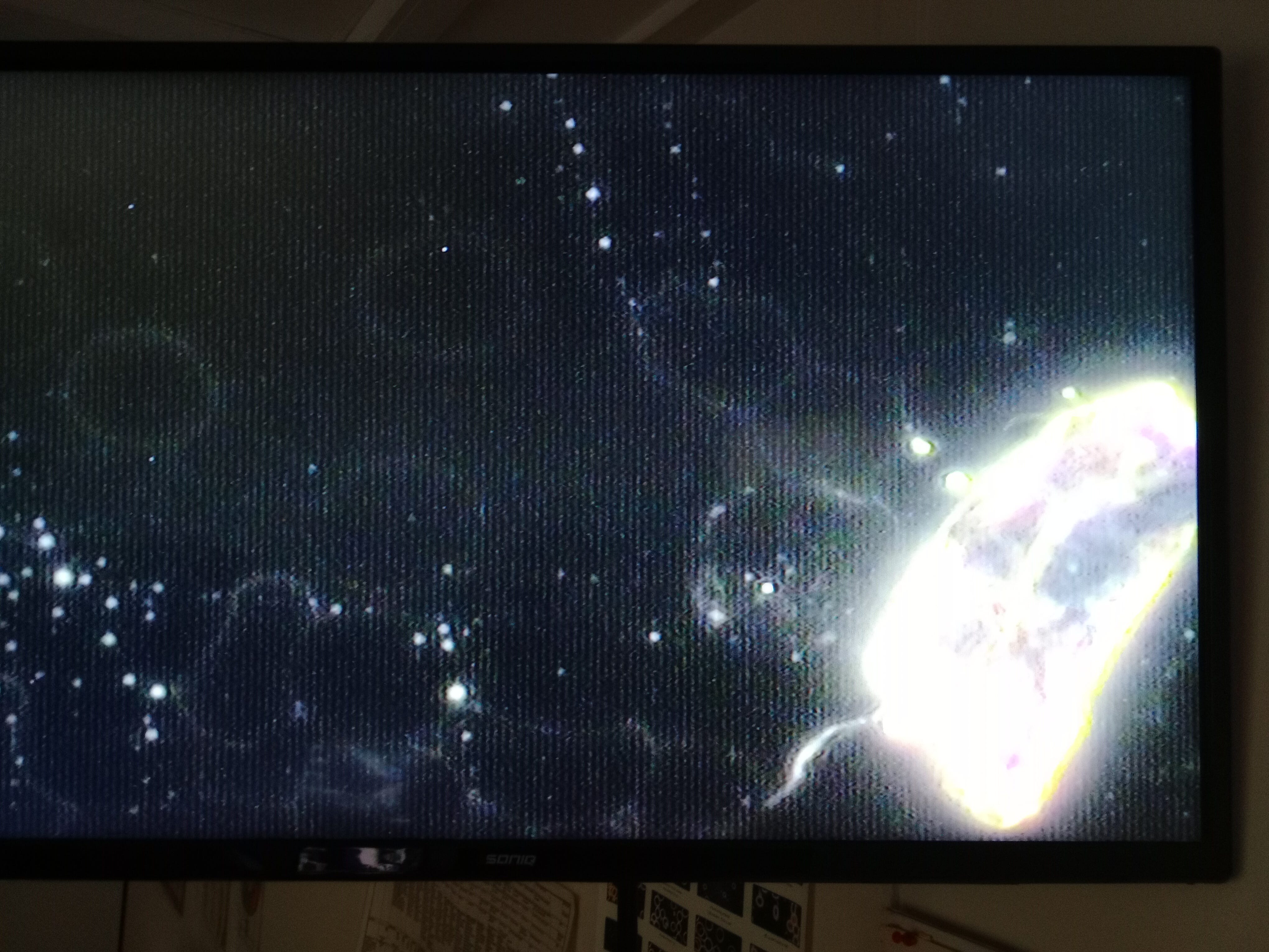
This is where I'll introduce a spray of similar filaments, as before completely new to us both.

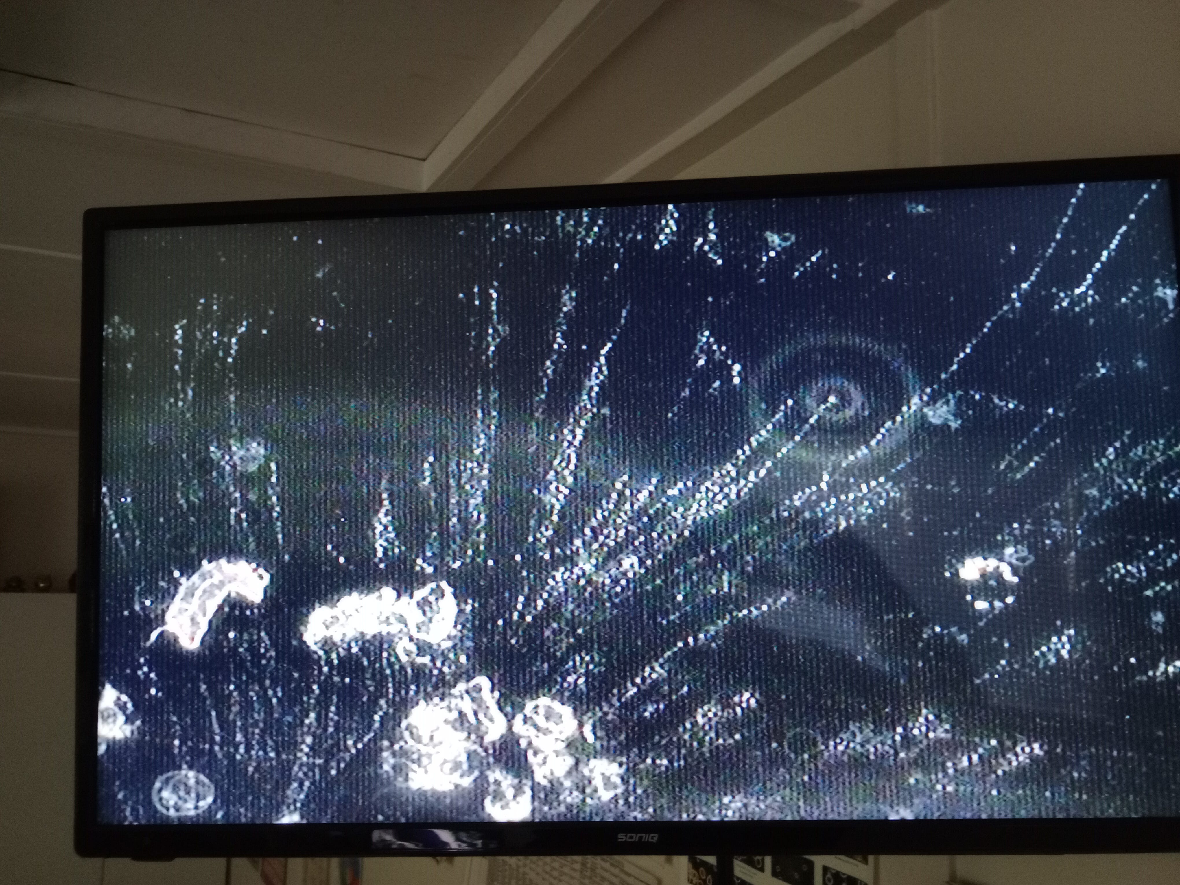
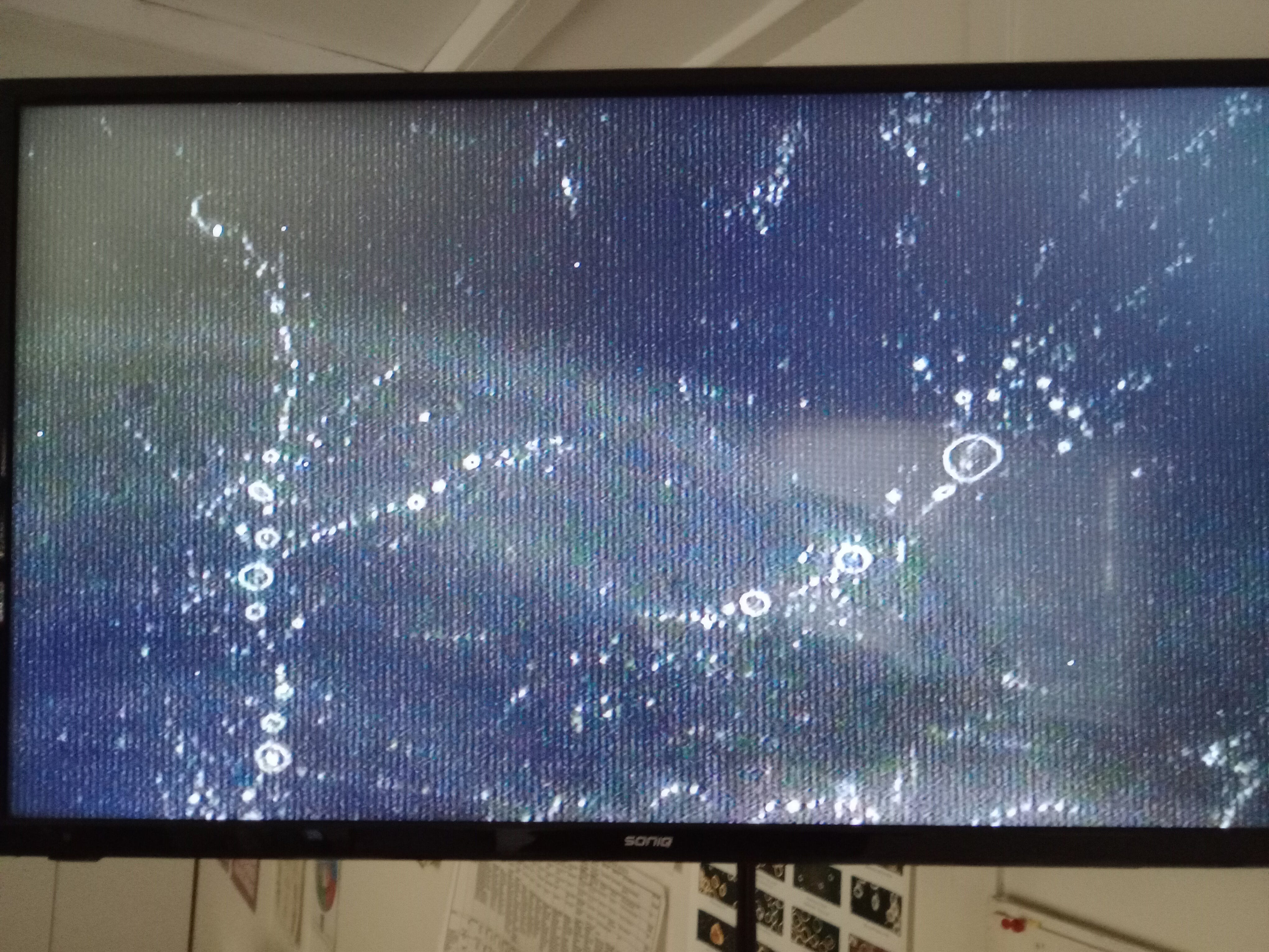
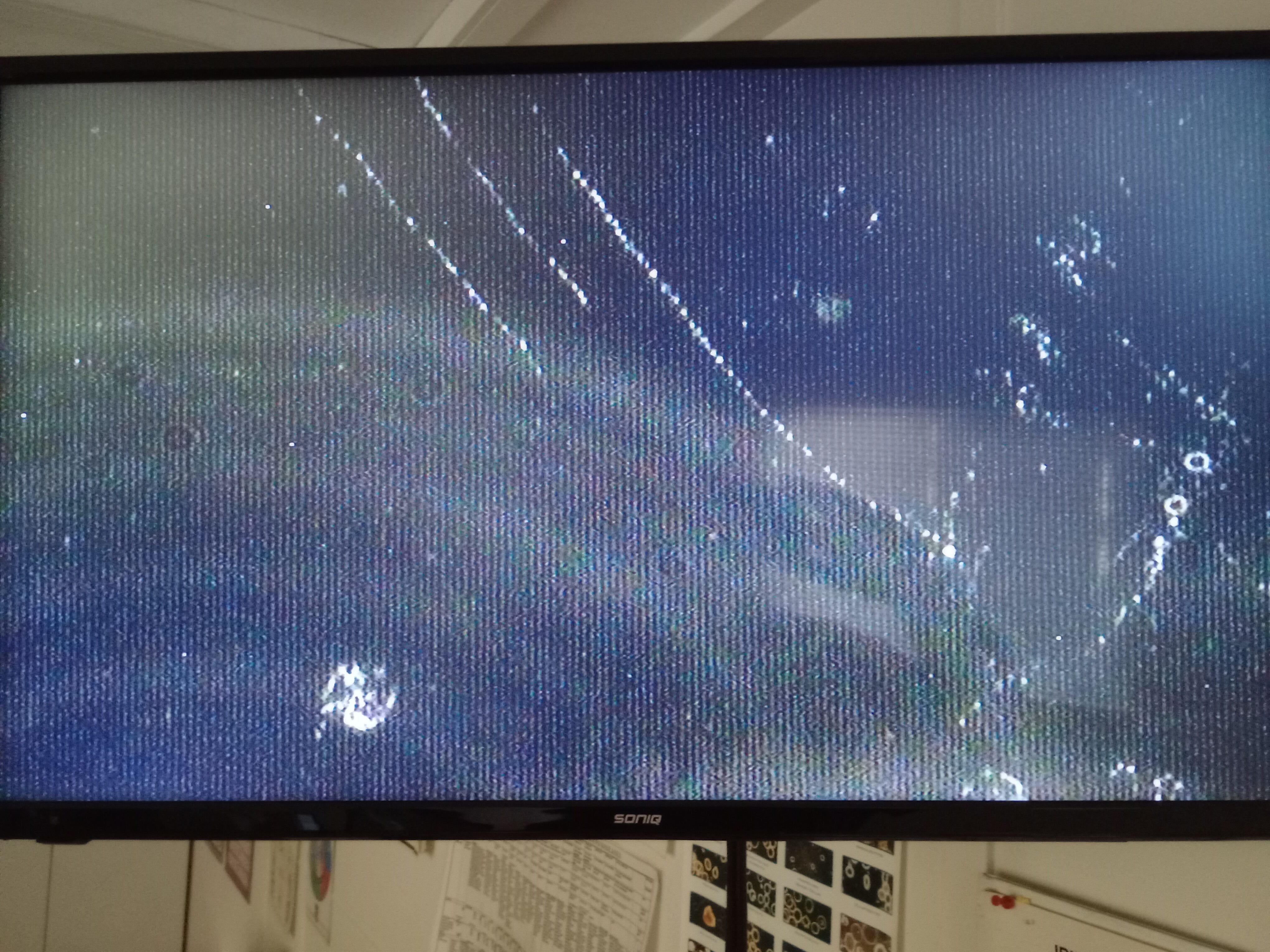
We tried to follow this spray
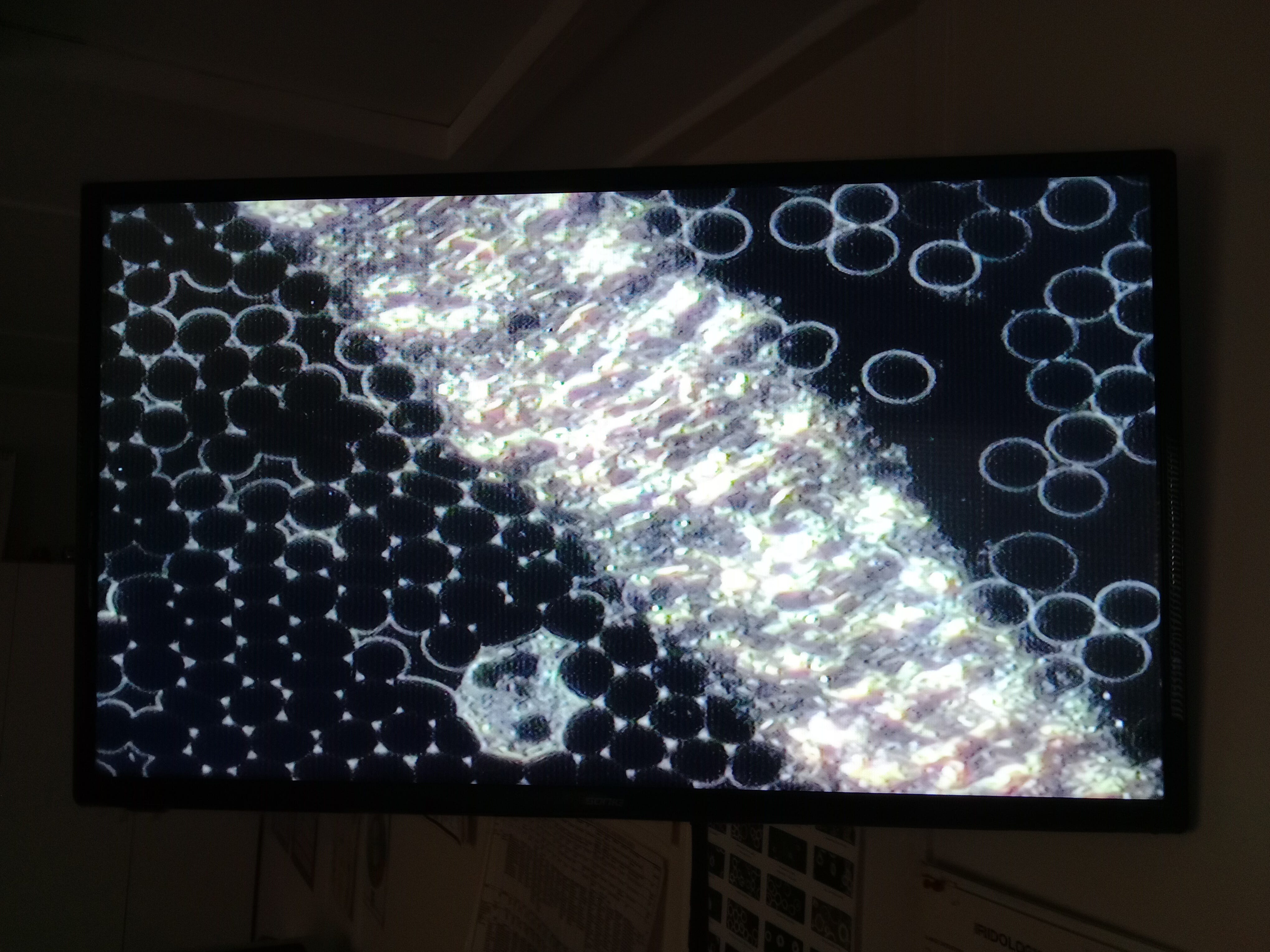
Still getting the shedding emptying itself and hopefully the photos are helpful down the track, if only to prove a crime.
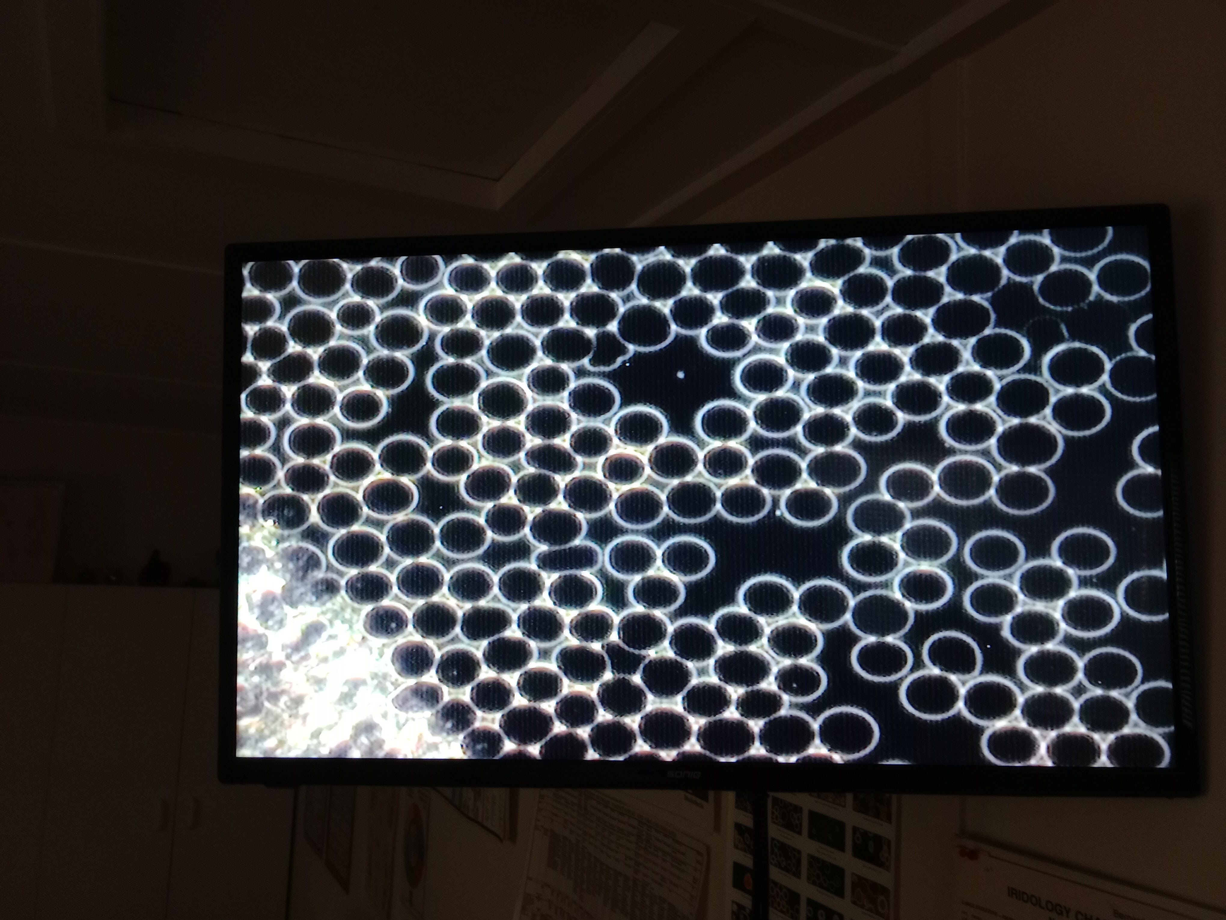
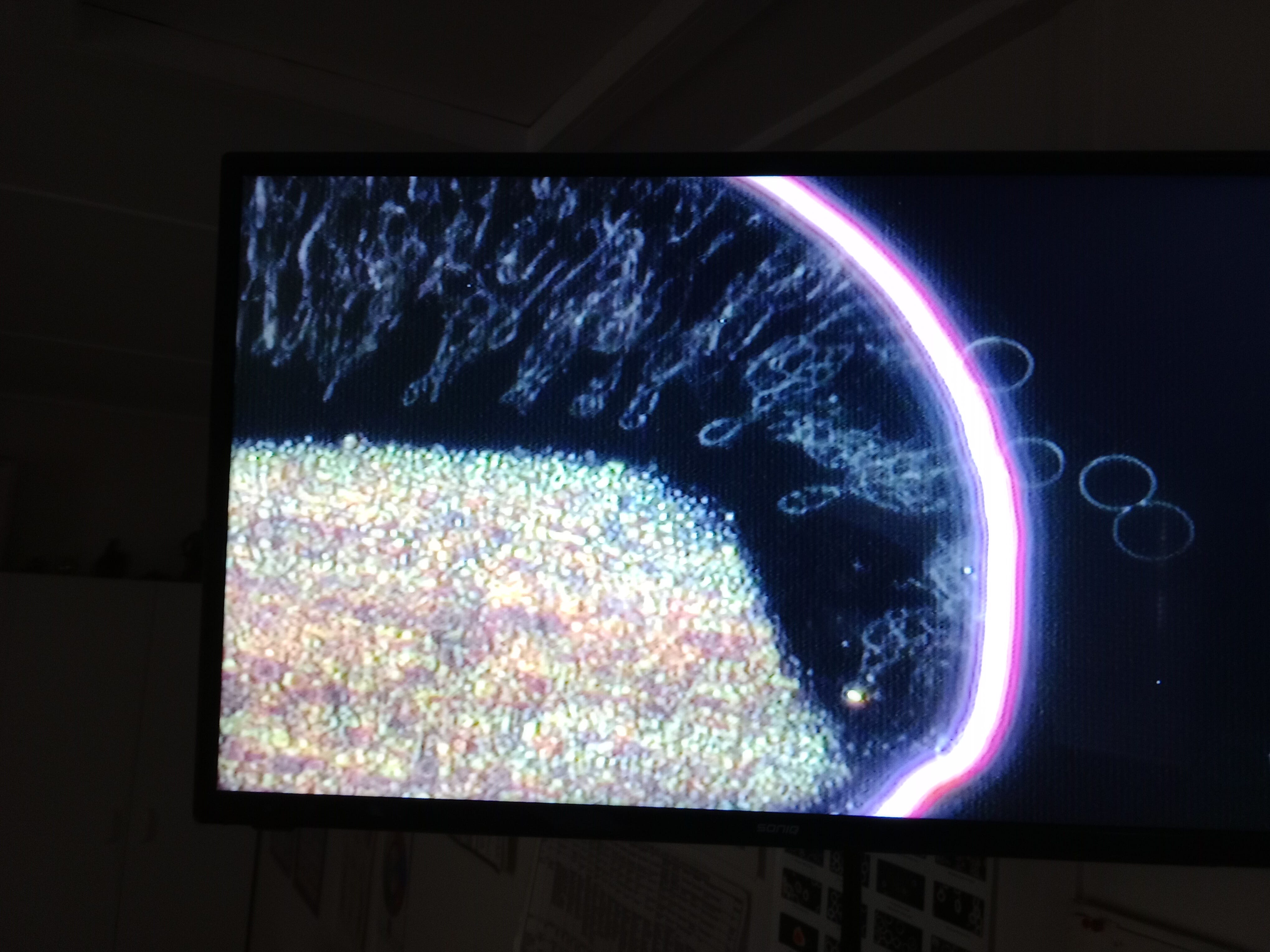
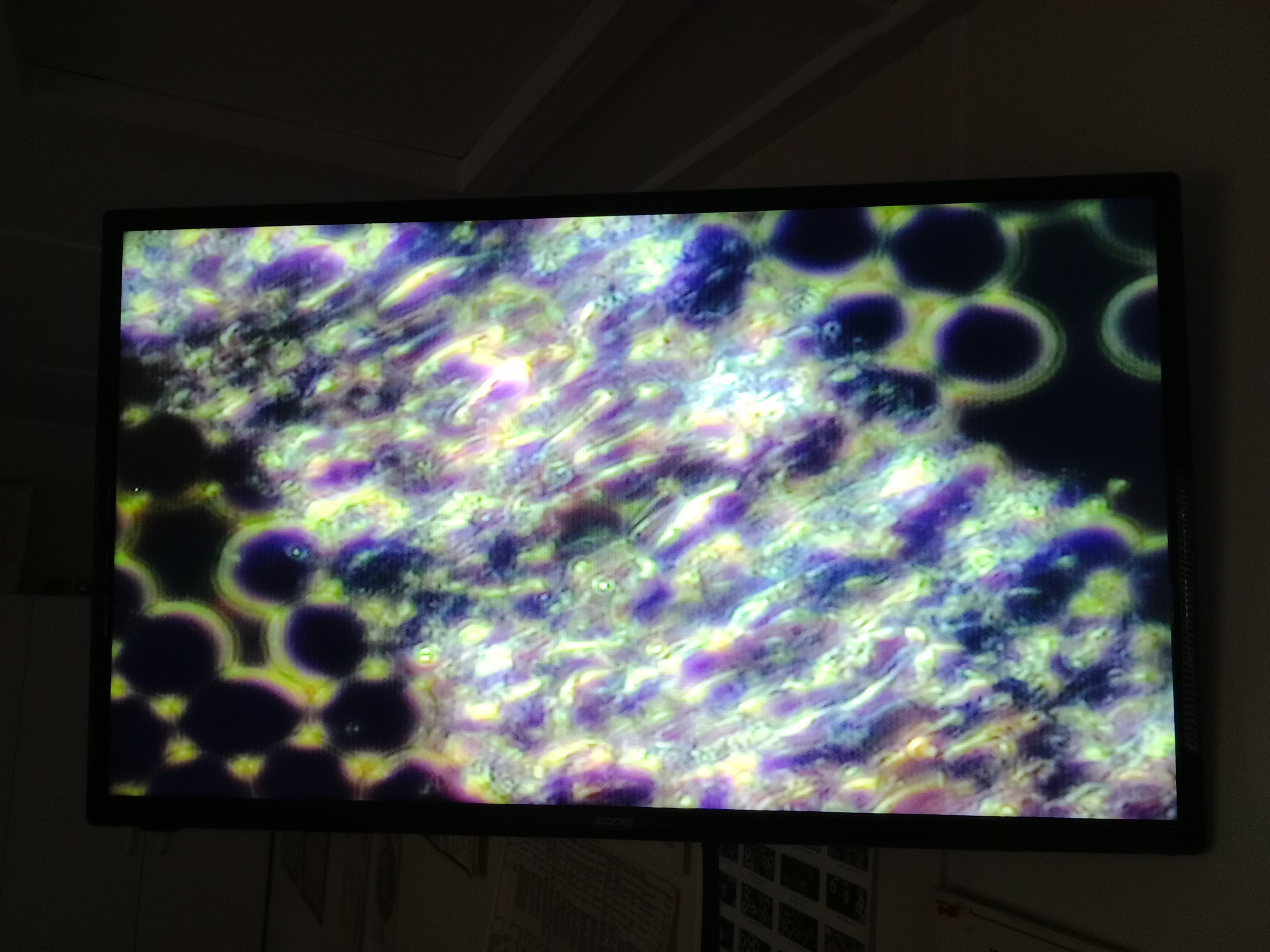
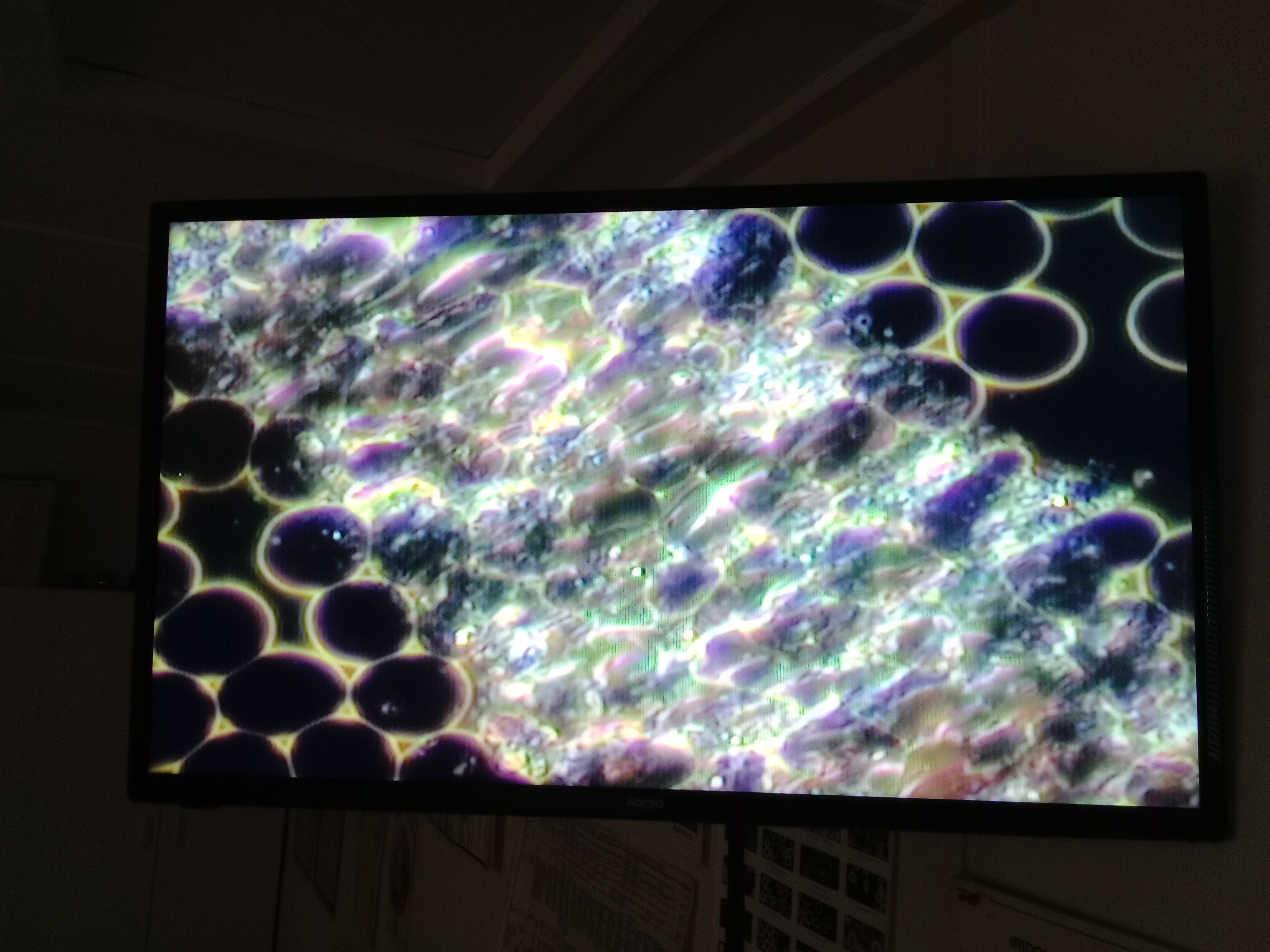
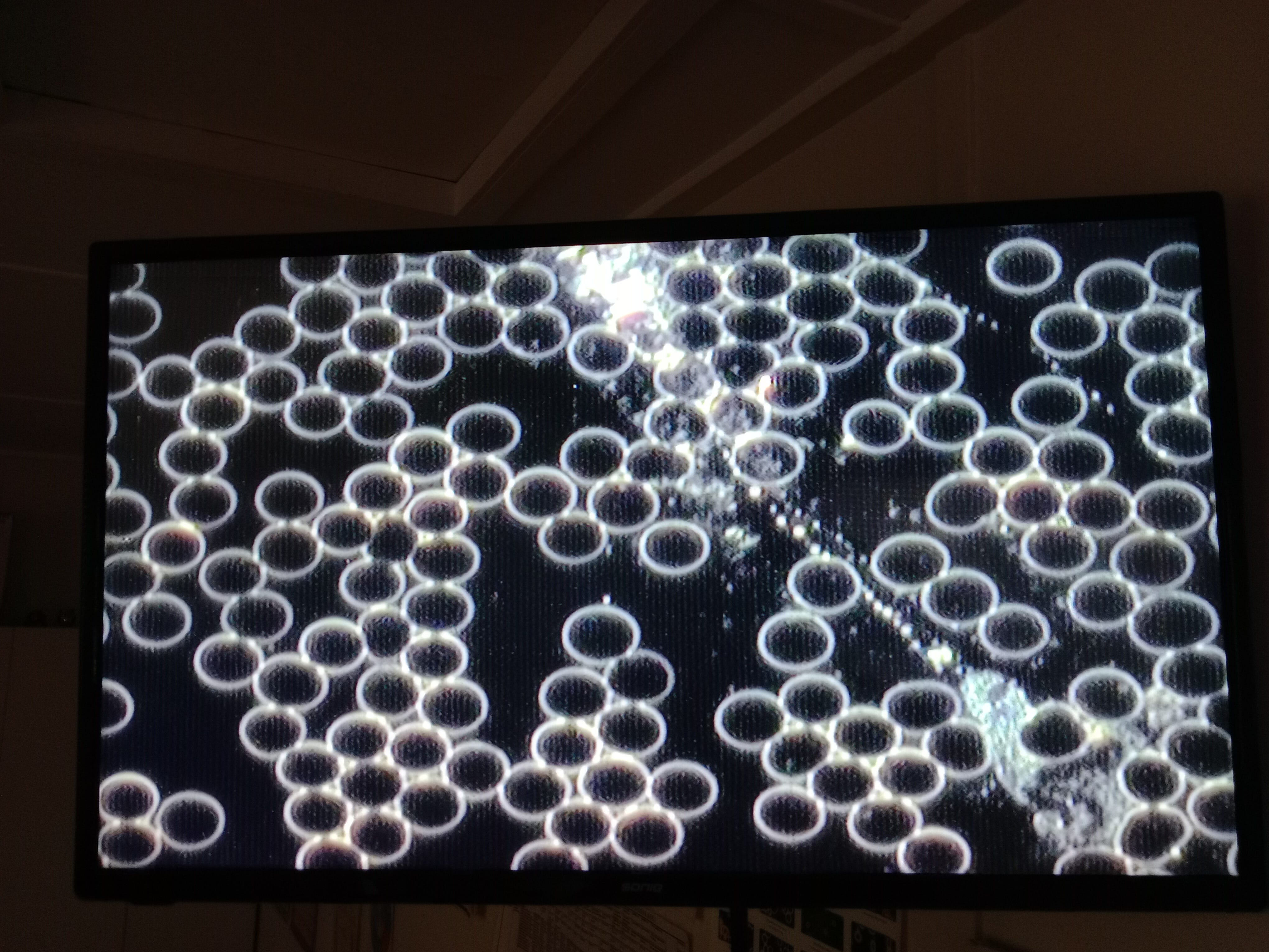
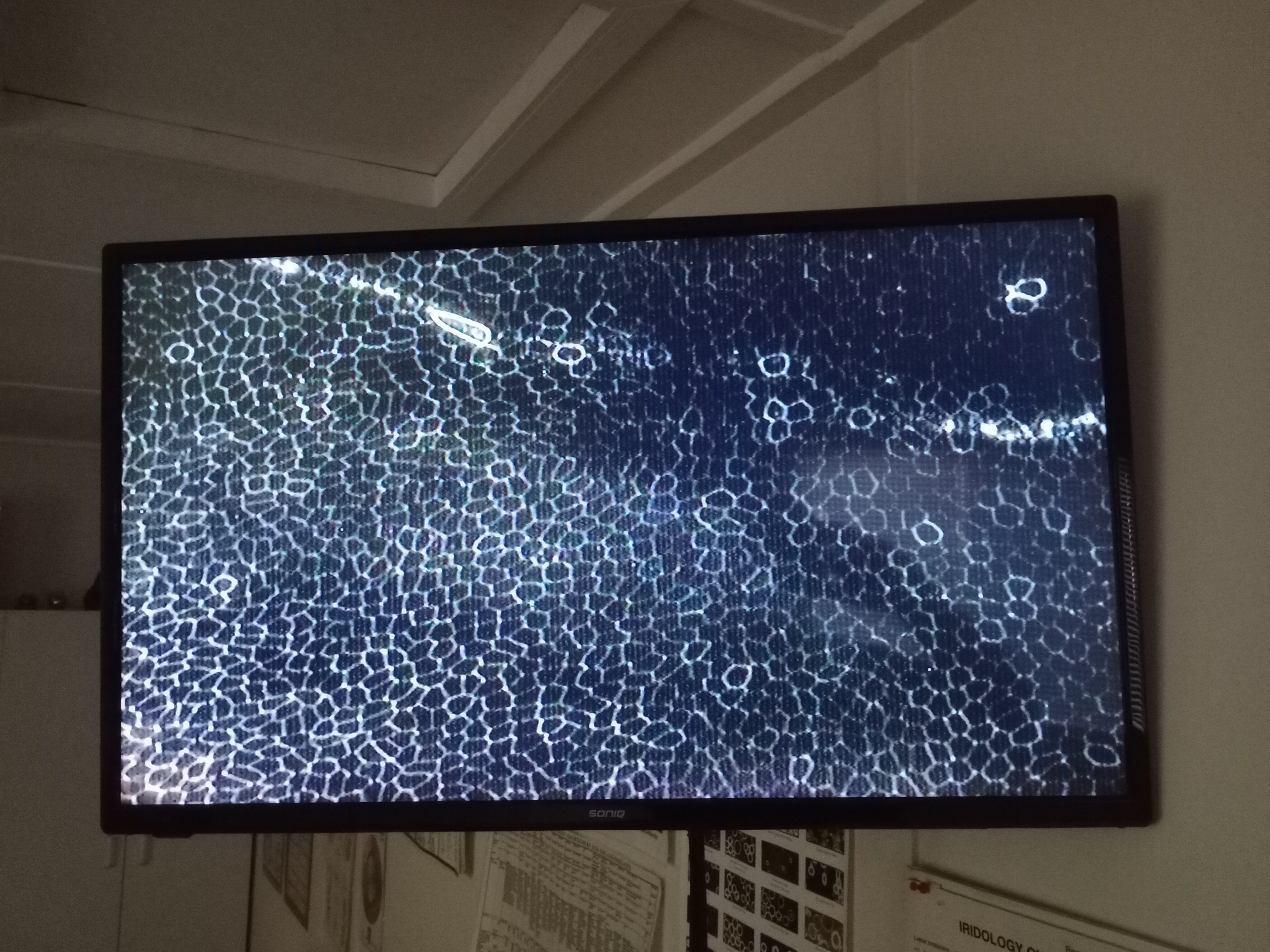
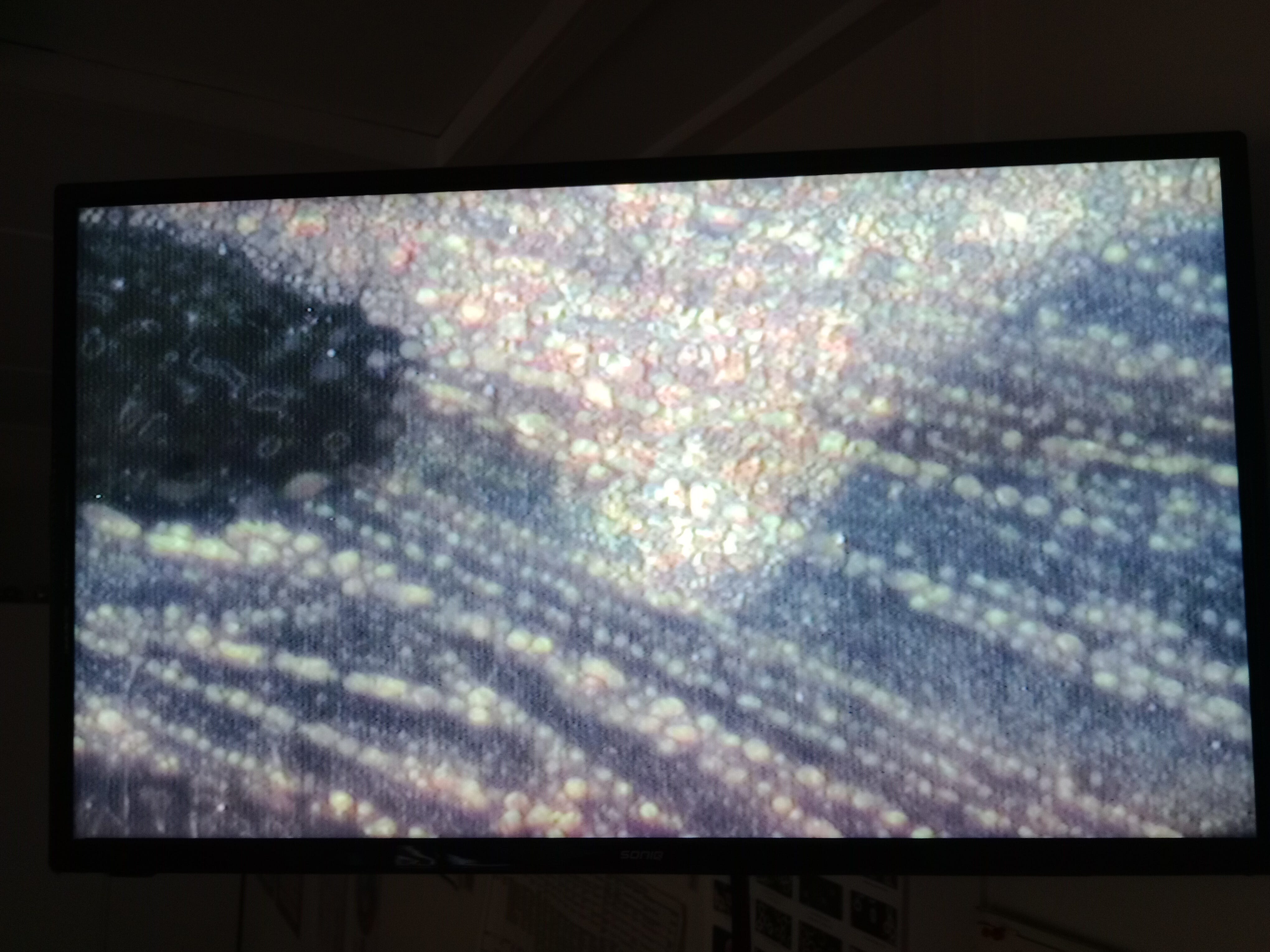
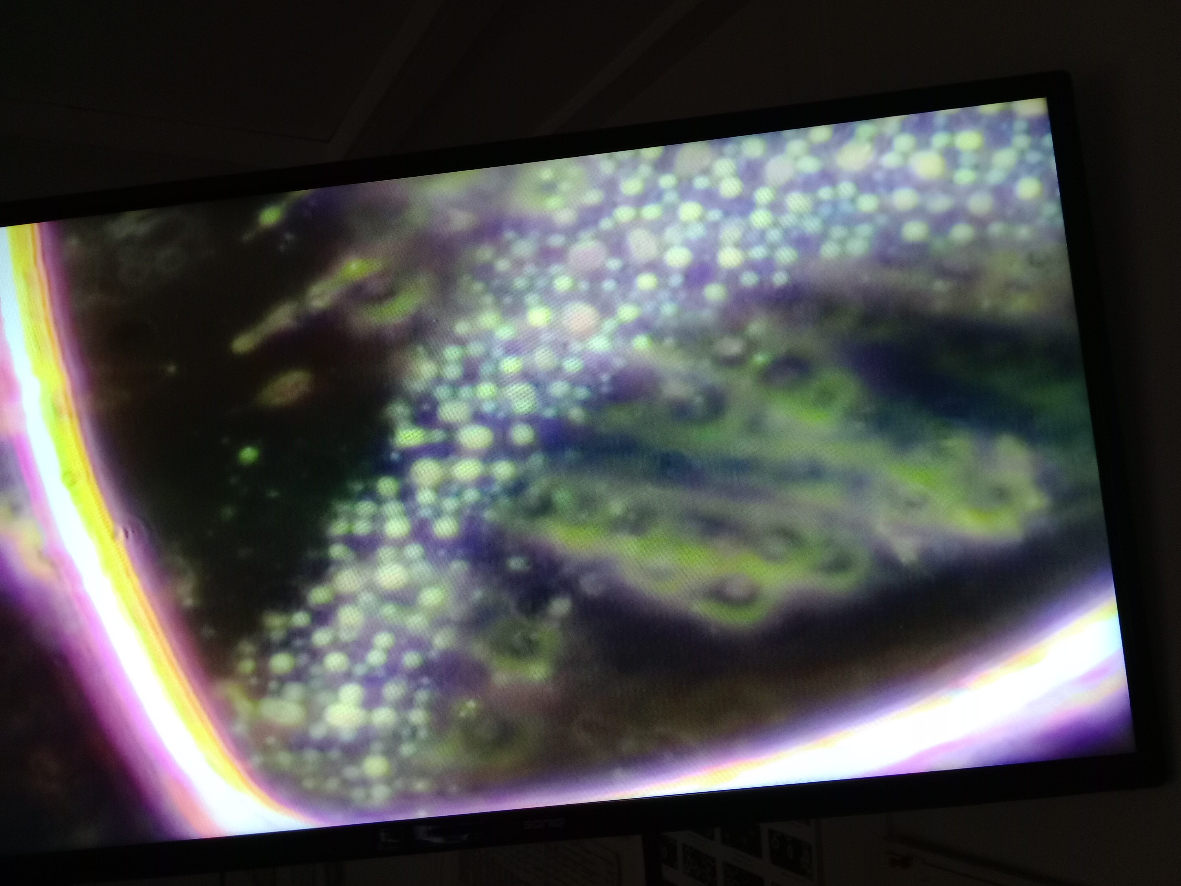
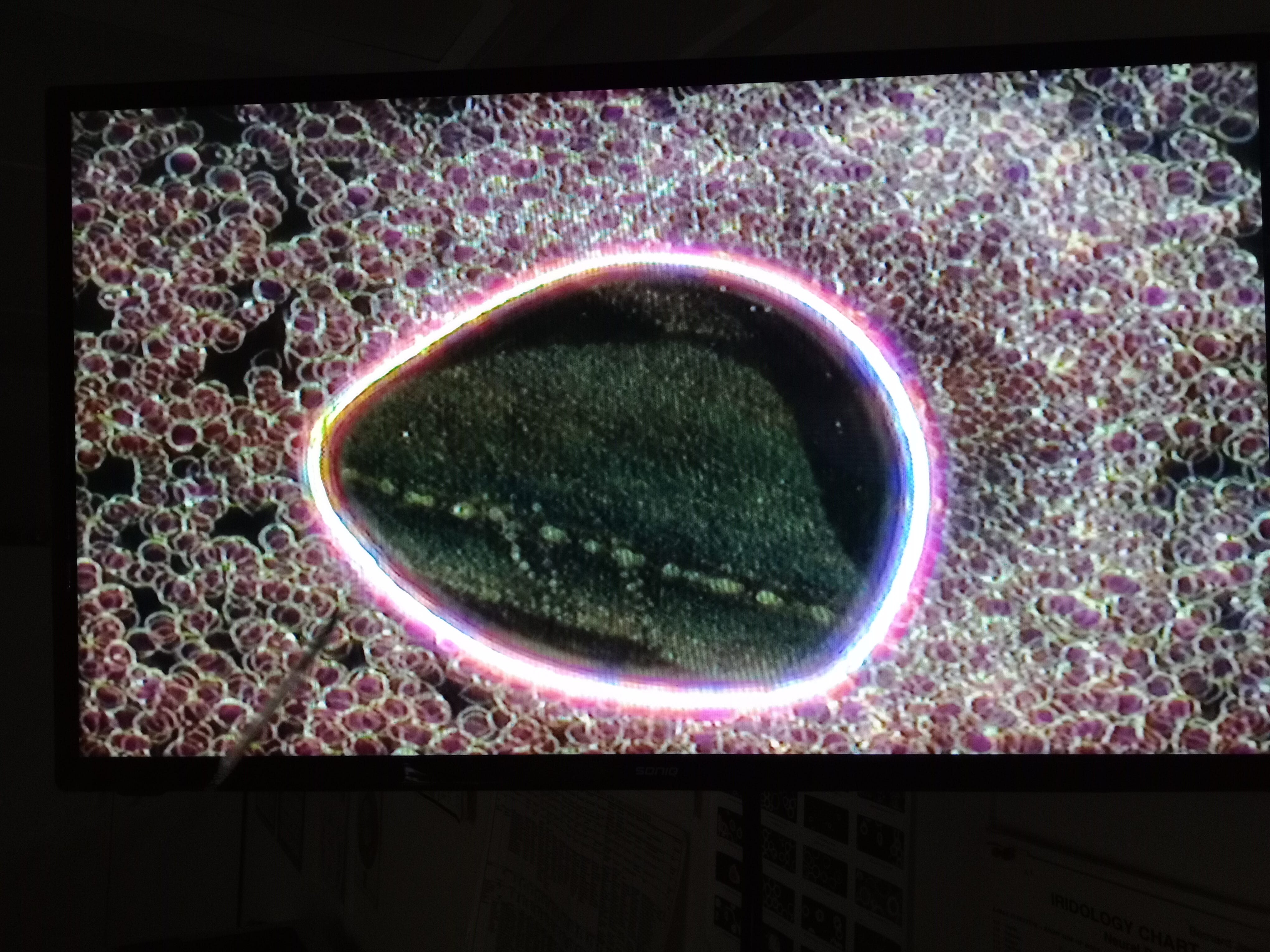
Before the shedding empty's out its fuller and the internal “particle” size is finer. It appears to lose the smaller particles first.
i.e. an old full photo
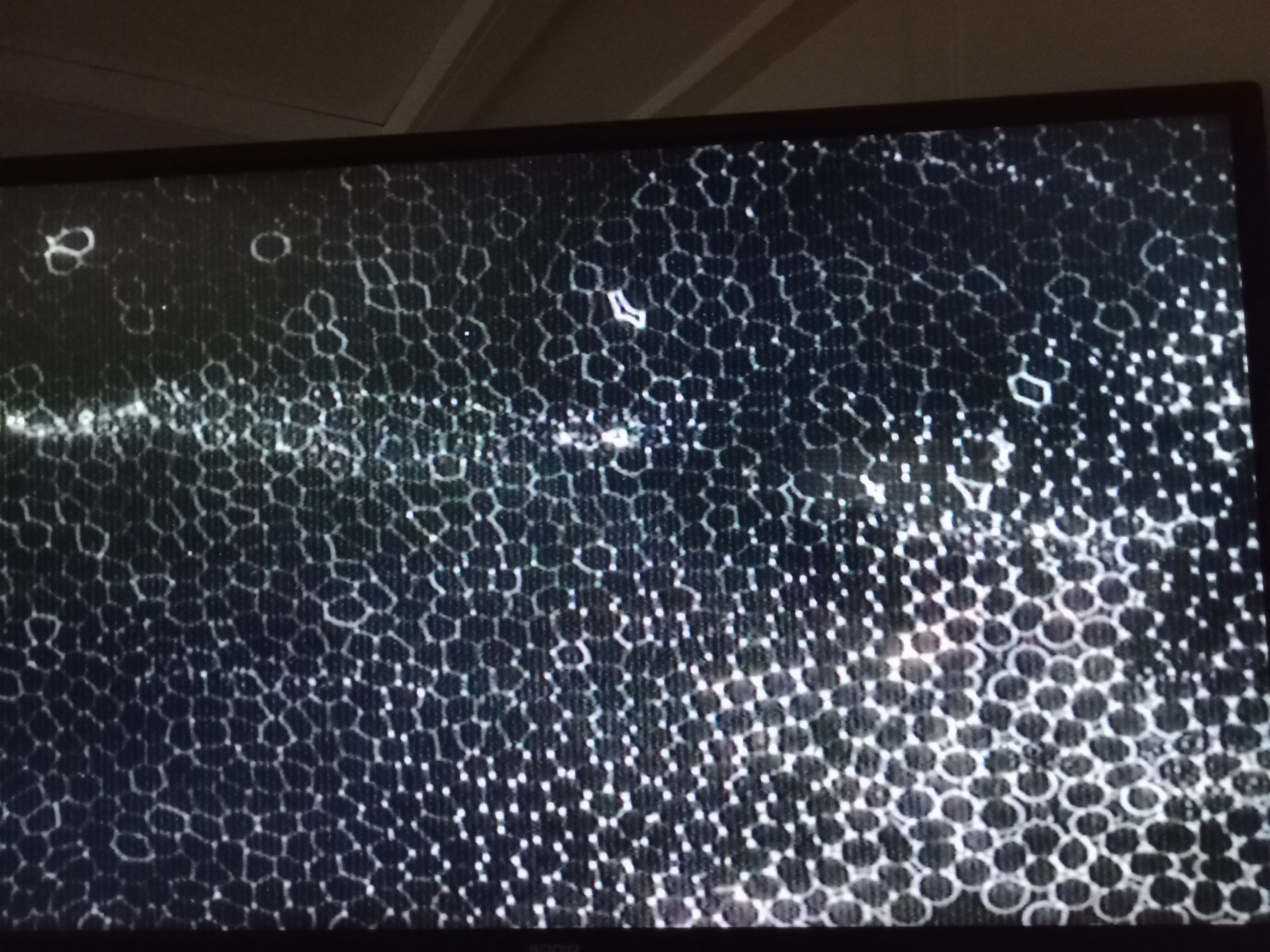
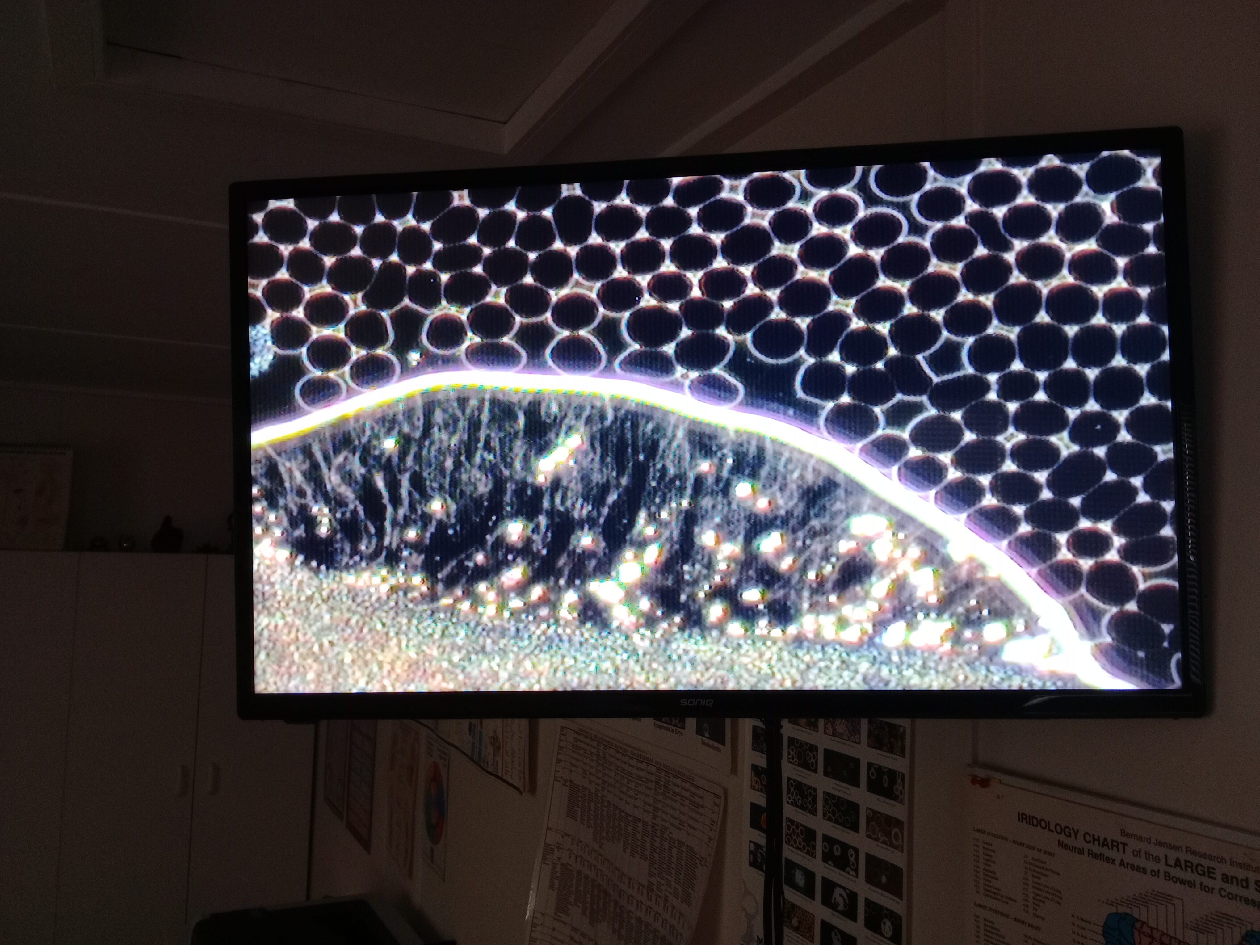
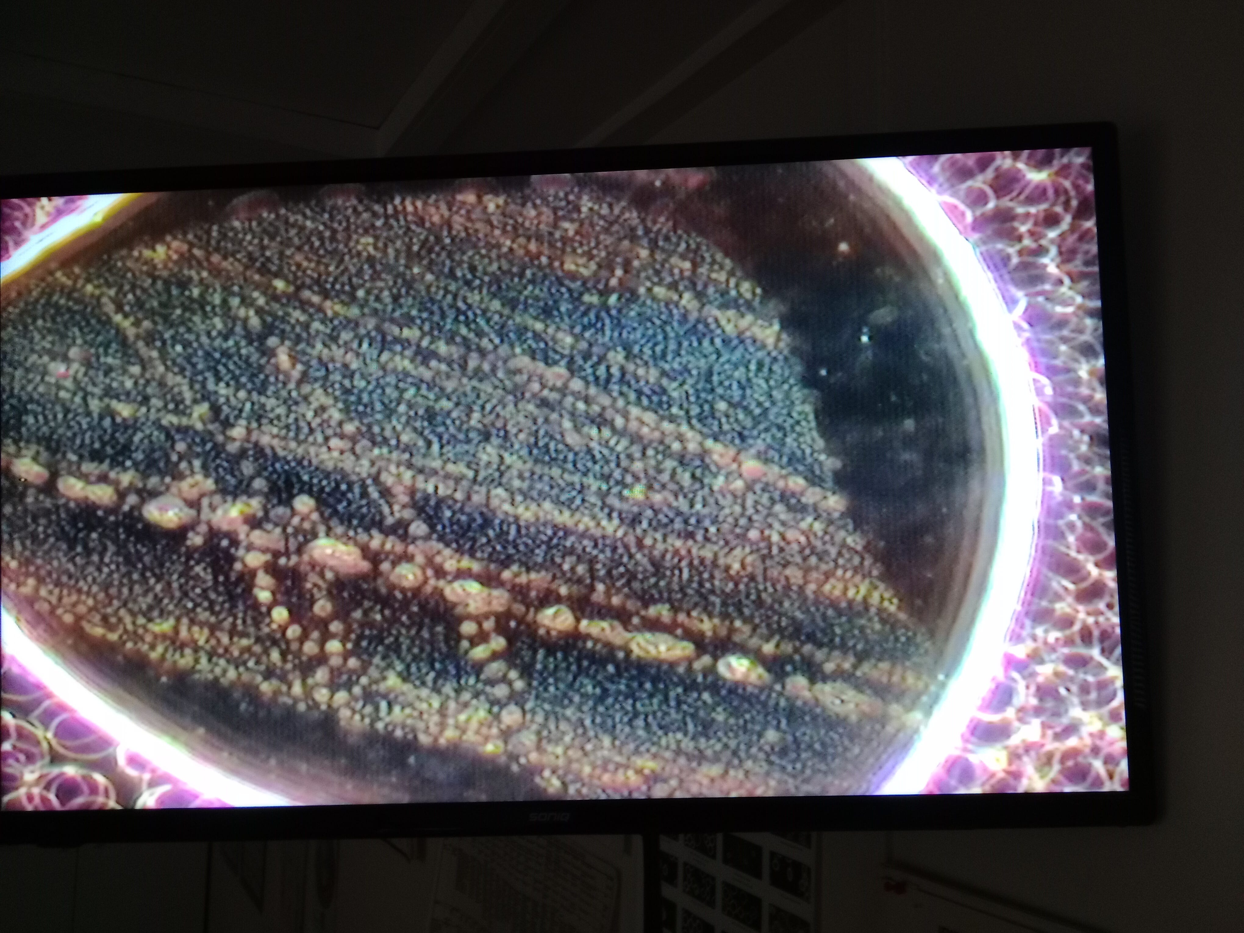
Compared to

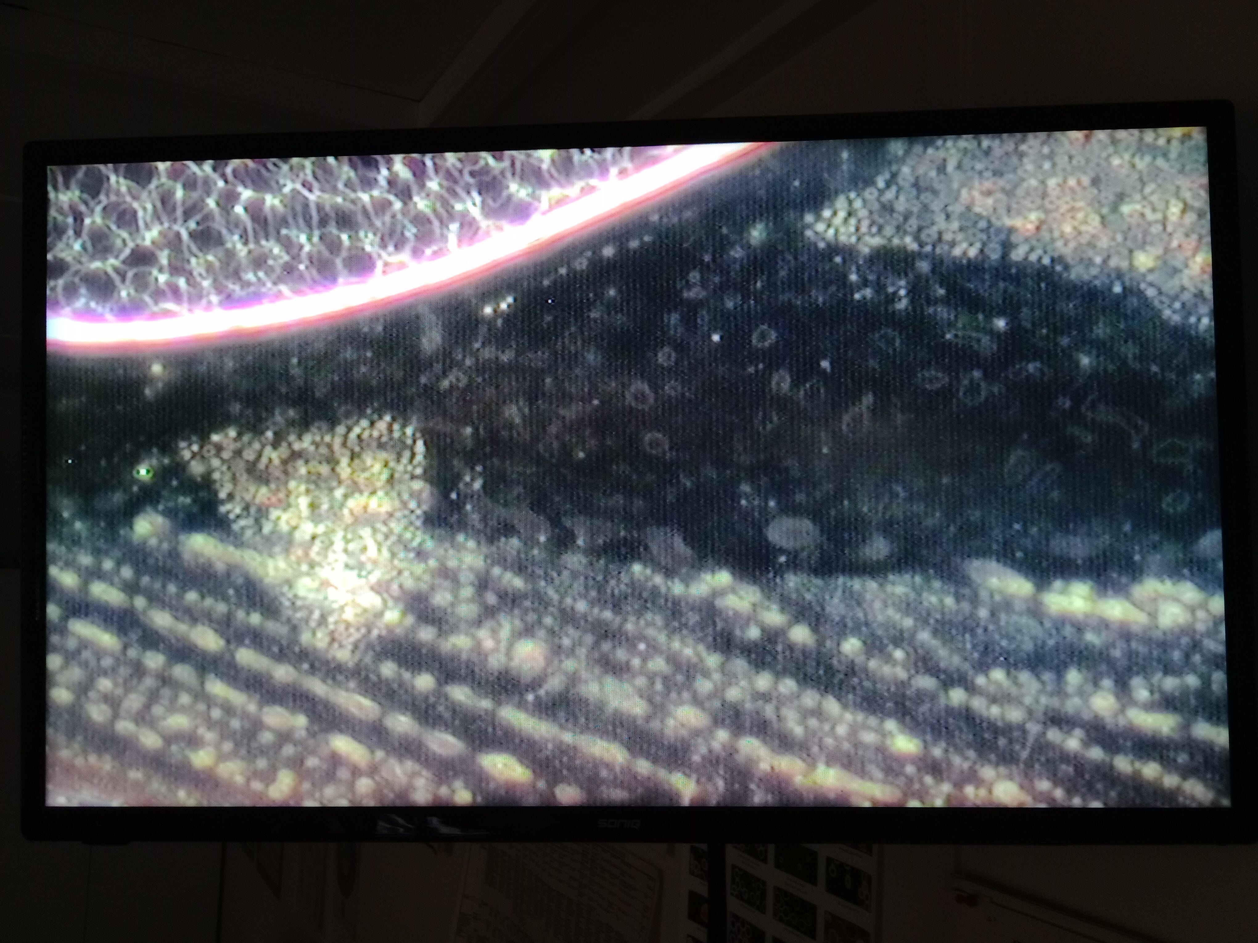
Whats interesting is that when it empty's out there appears to be a courser grain running in lines, these lines all appear to be running in the same direction even though the bubbles “appear” independent of each other. Perhaps it is a frequency based primer and like a wave flowing through us they all activate in line with each other.
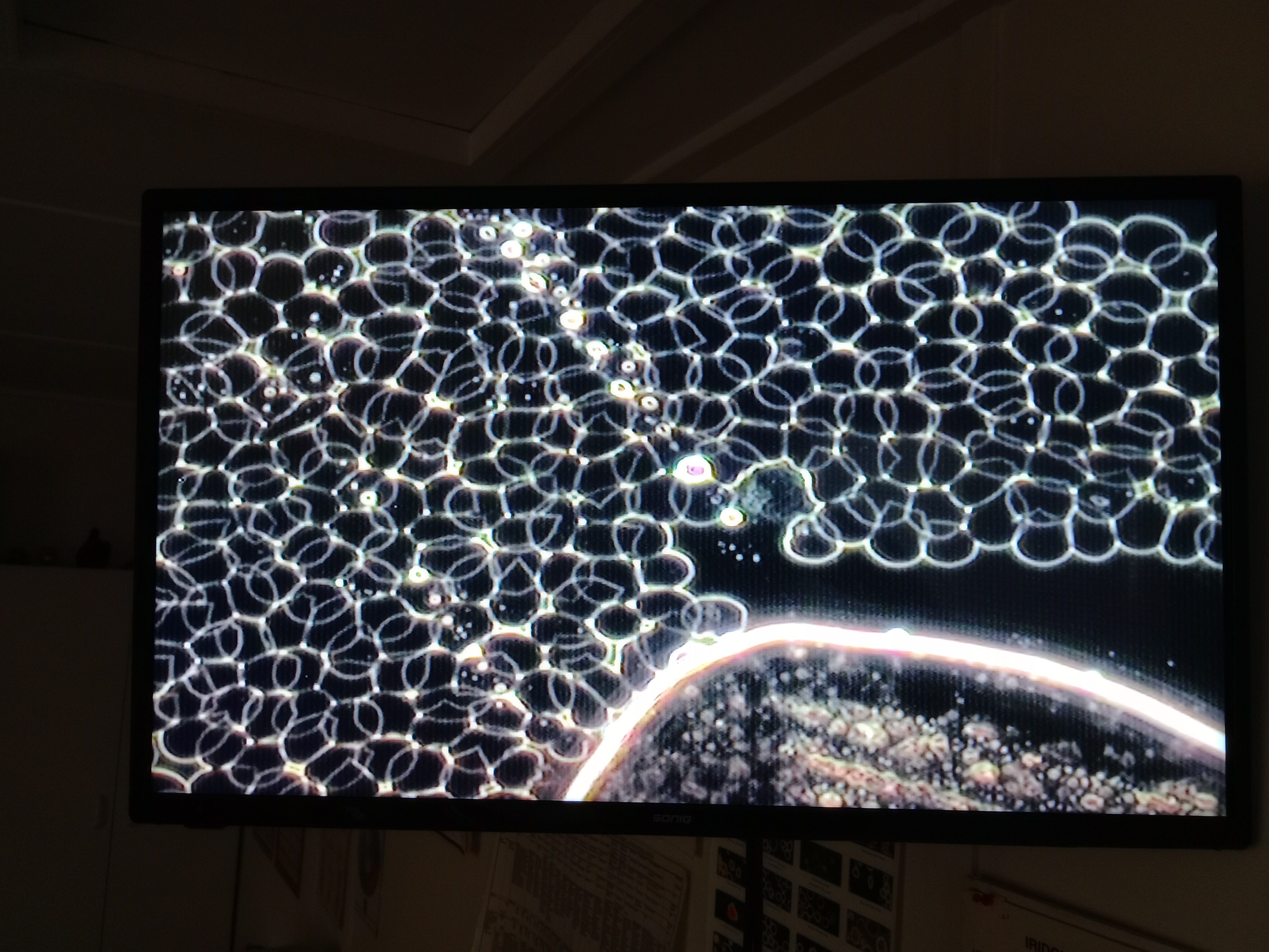
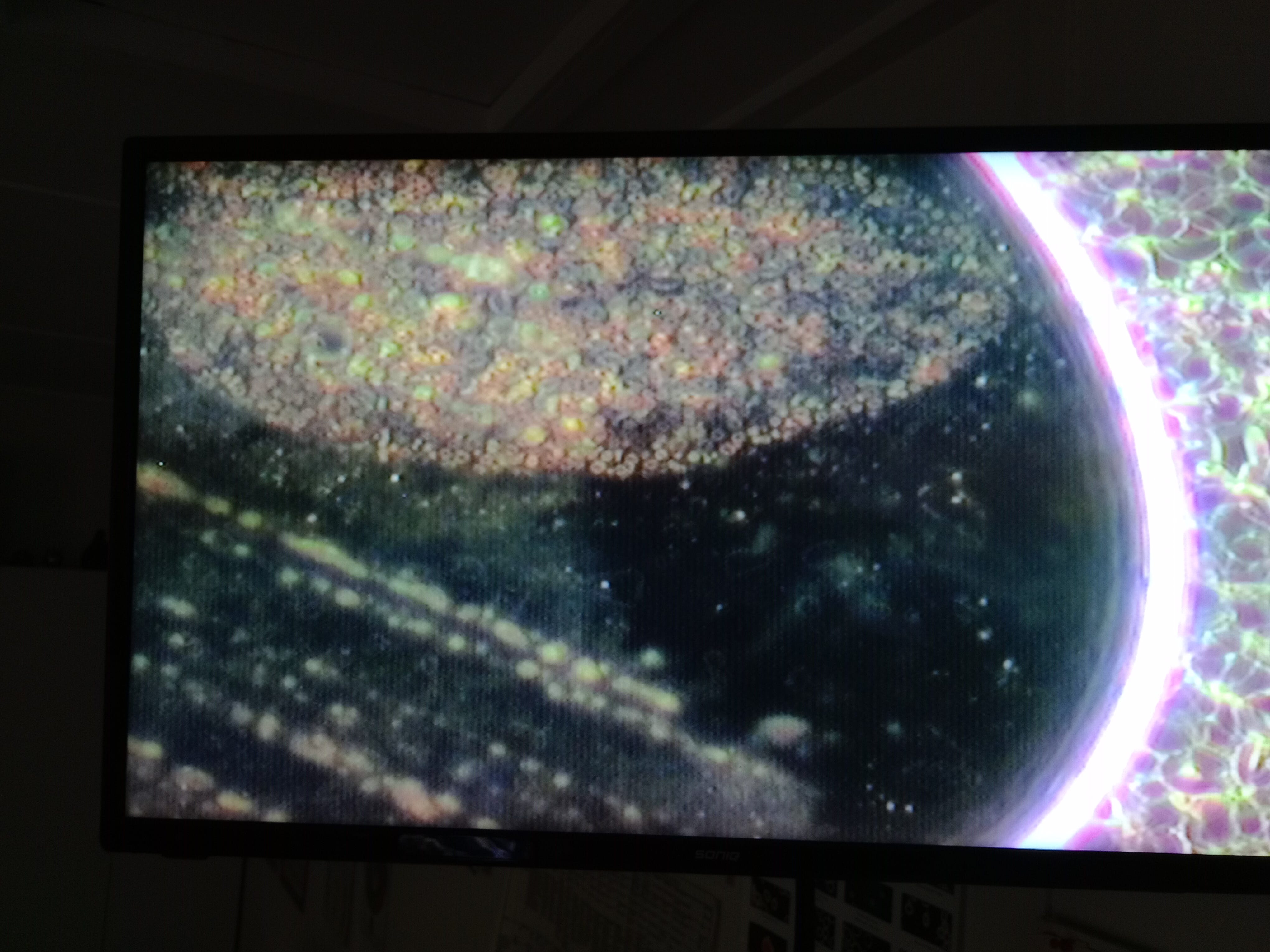
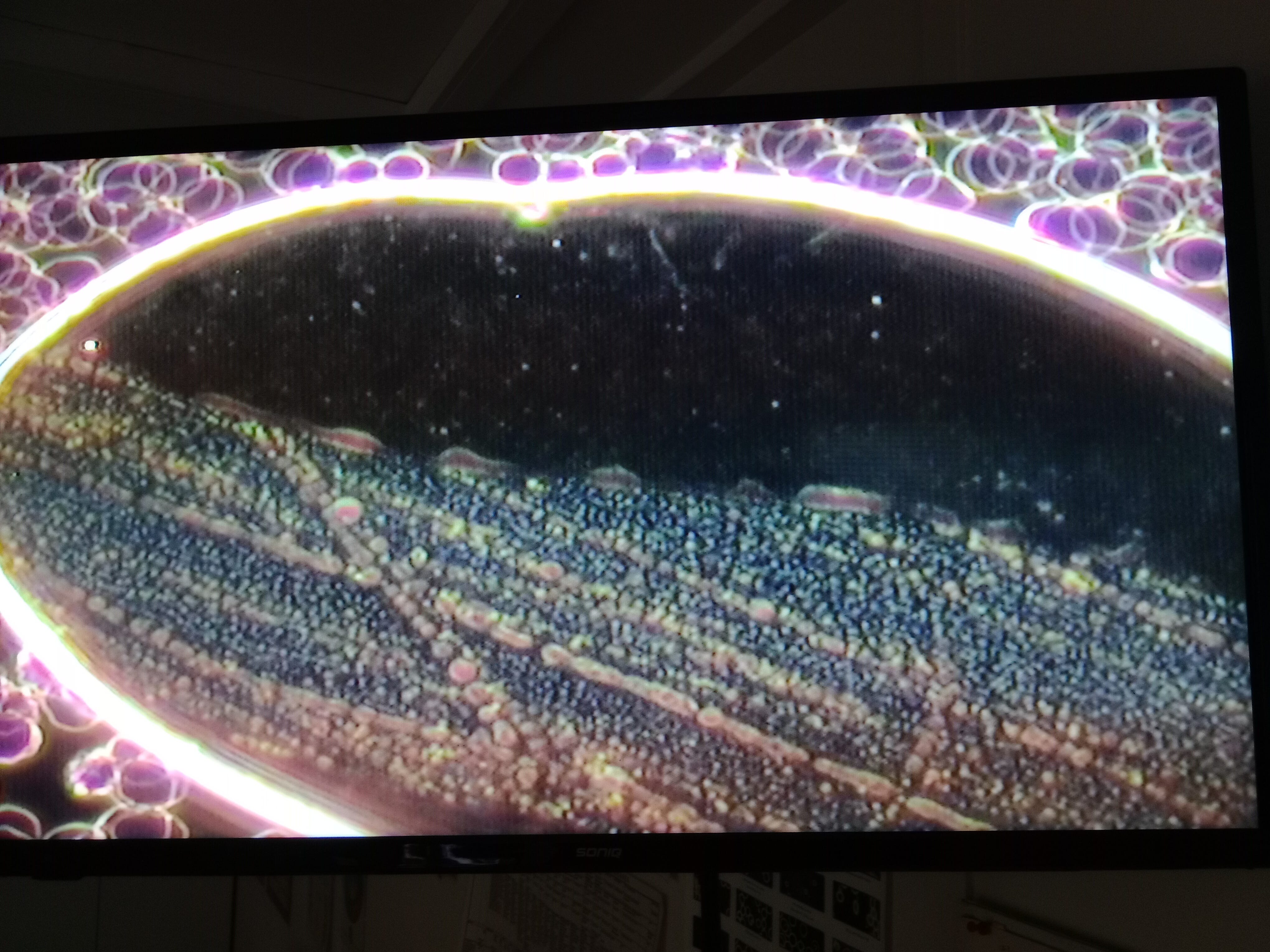
We slowly change the focus to potentially show depth/levels
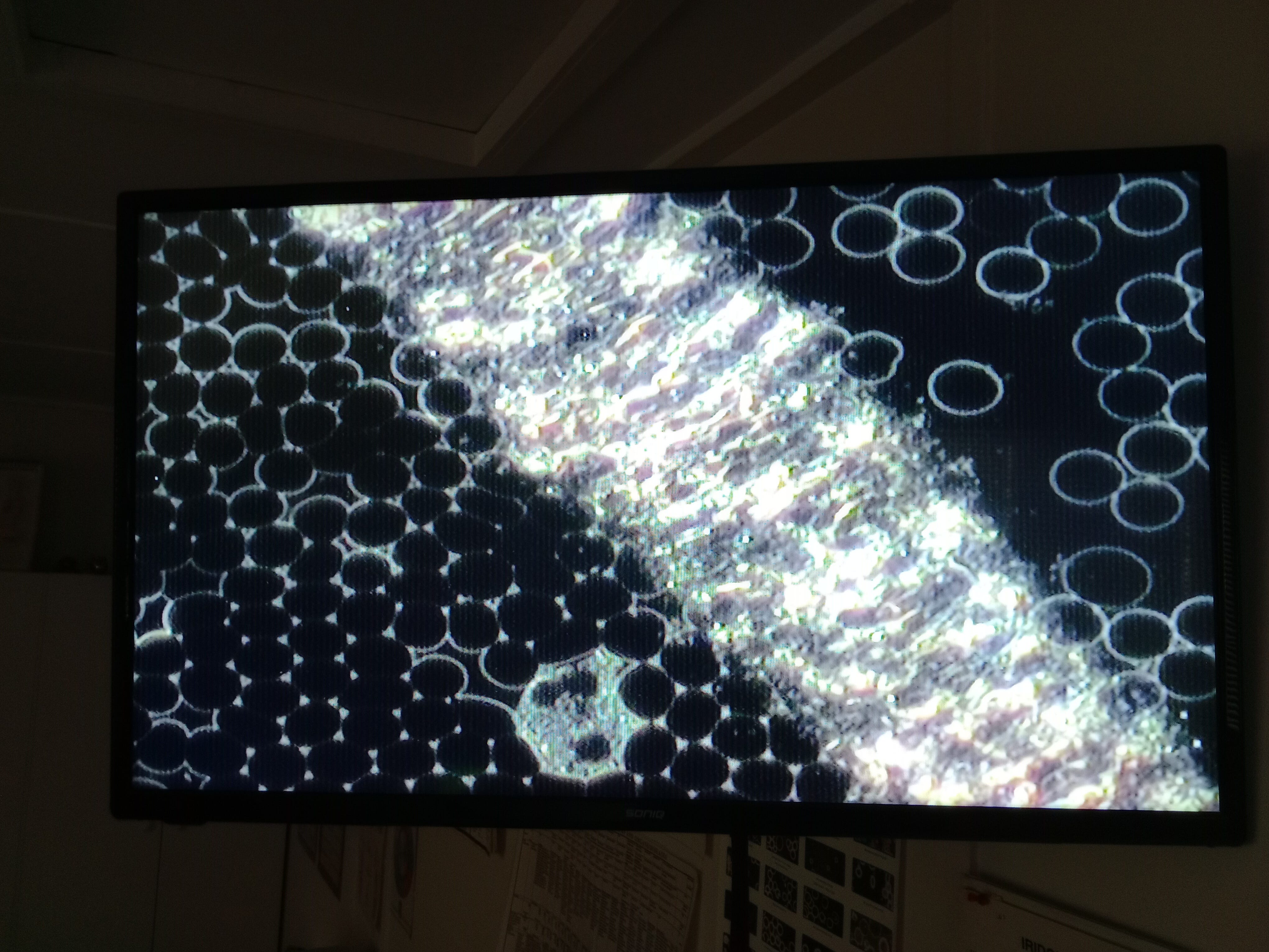
If we look at the following there is an underlying pattern, almost like soil packed down over the years.
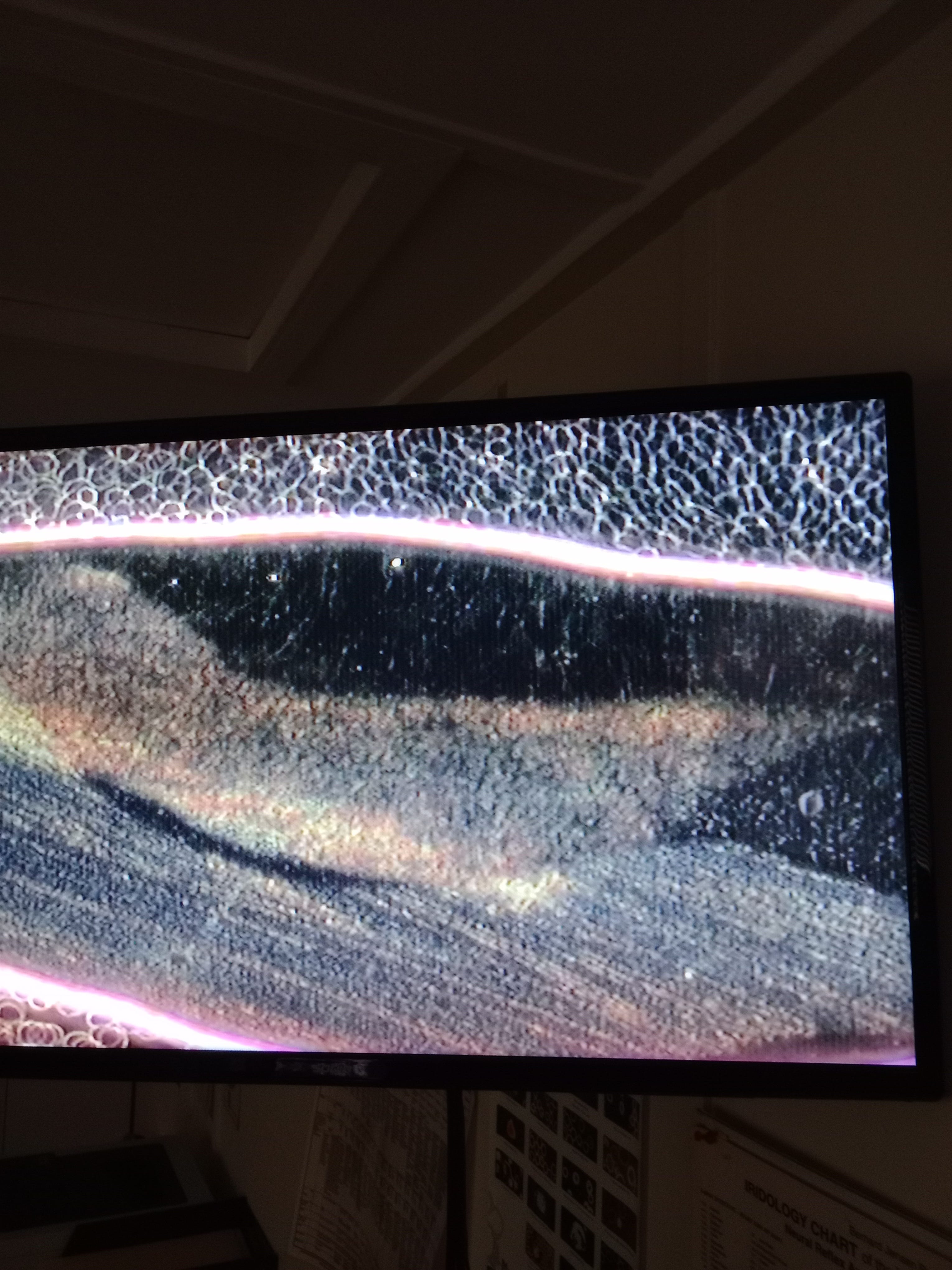

And this one as well.
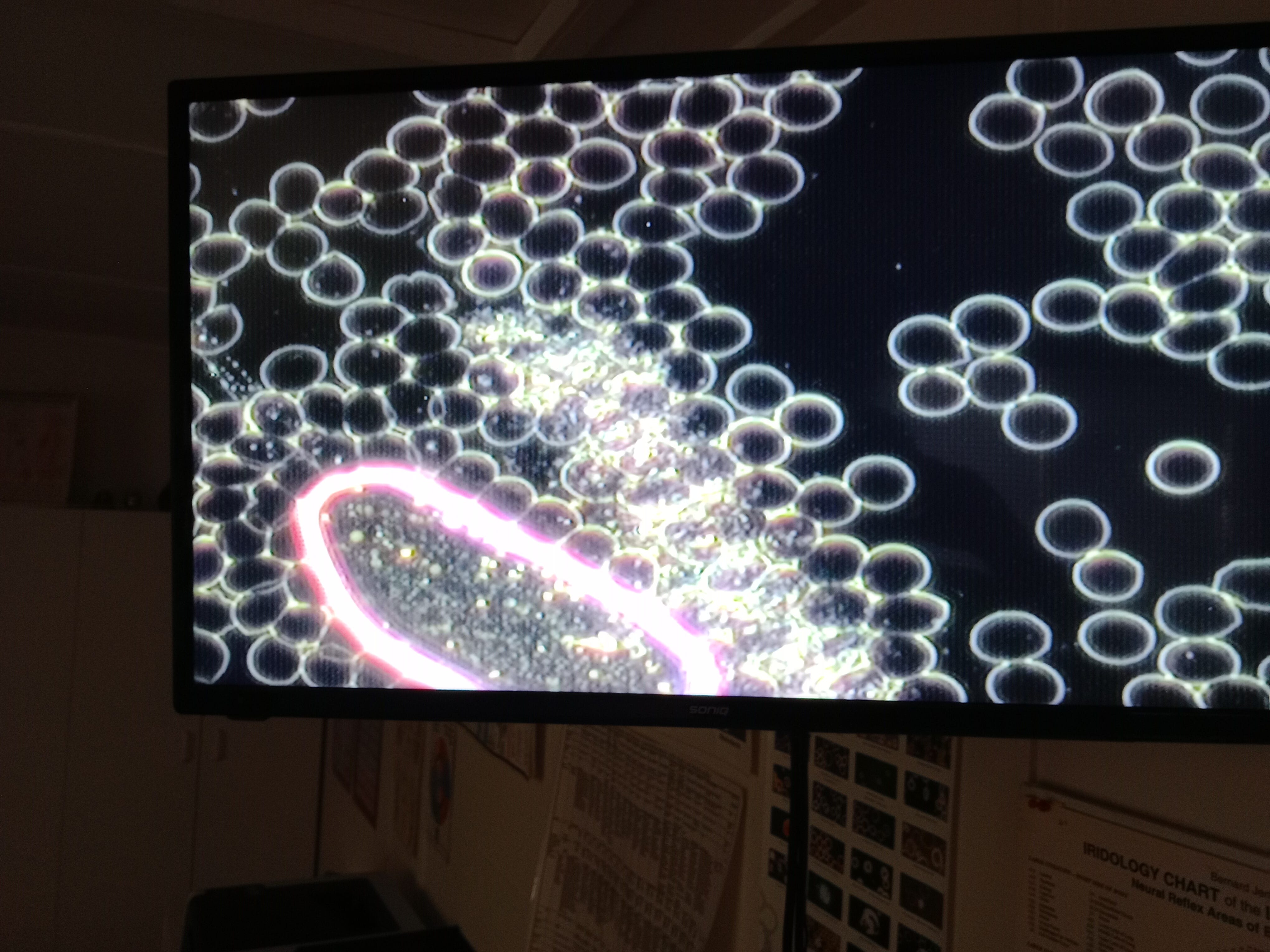
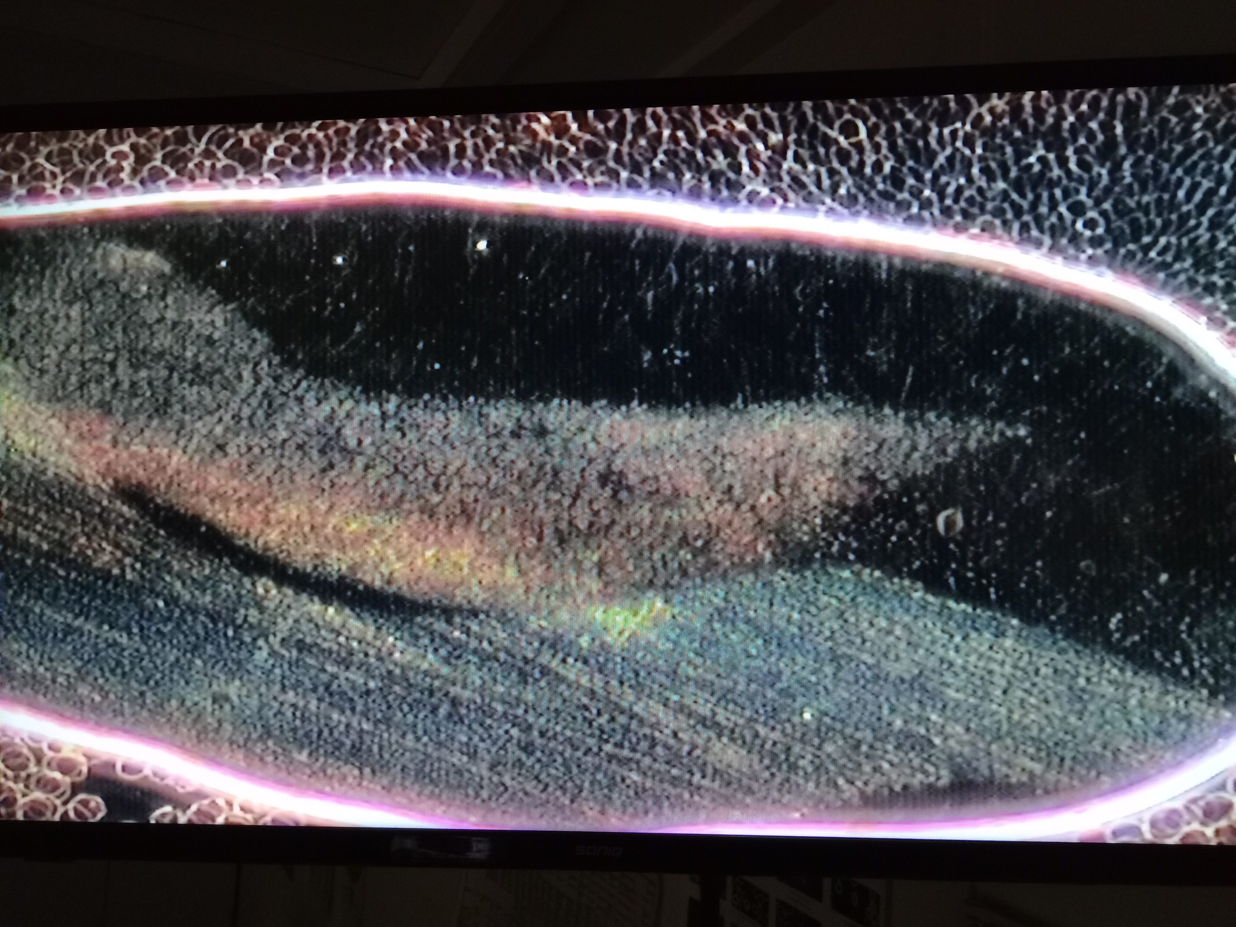
Another example of the clumping/clotting closest to the empty side BUT its not an absolute.
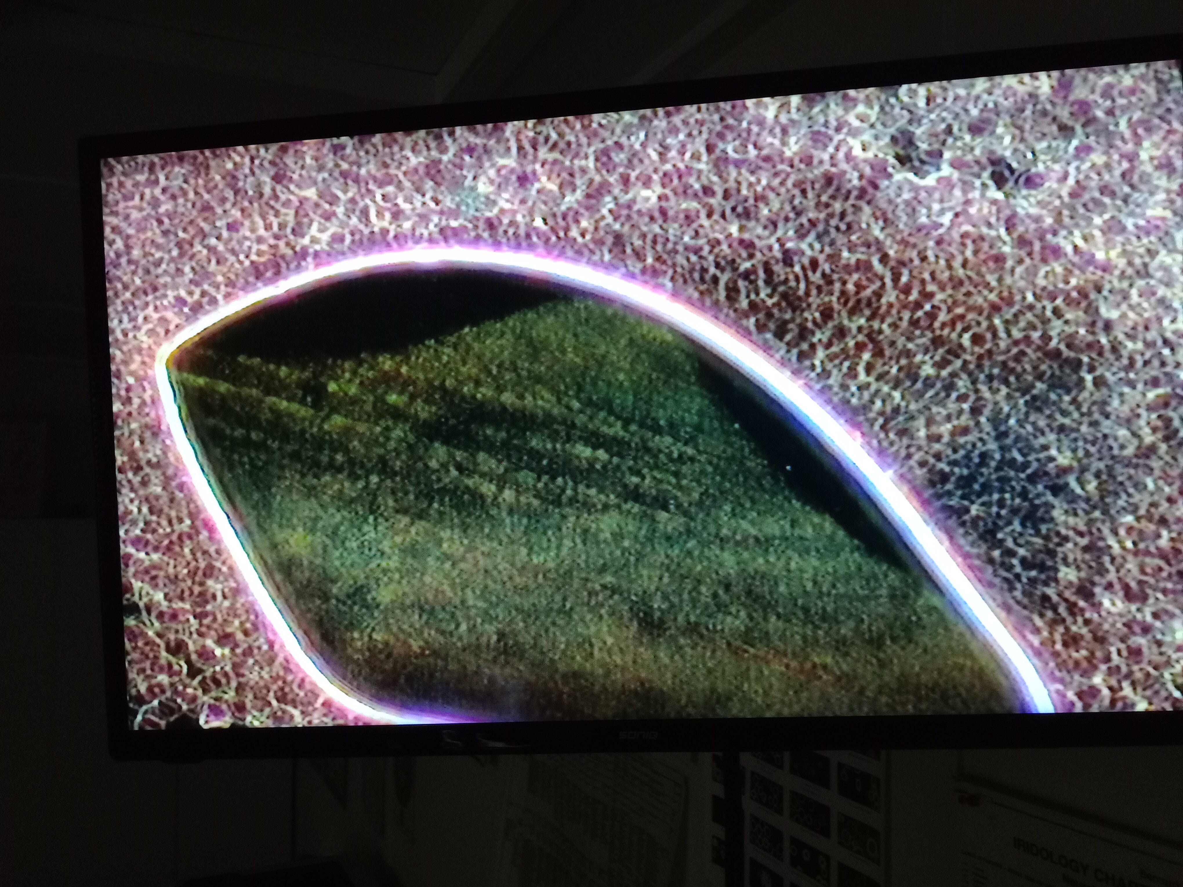
Go back a little and you have a better idea on cause and effect.
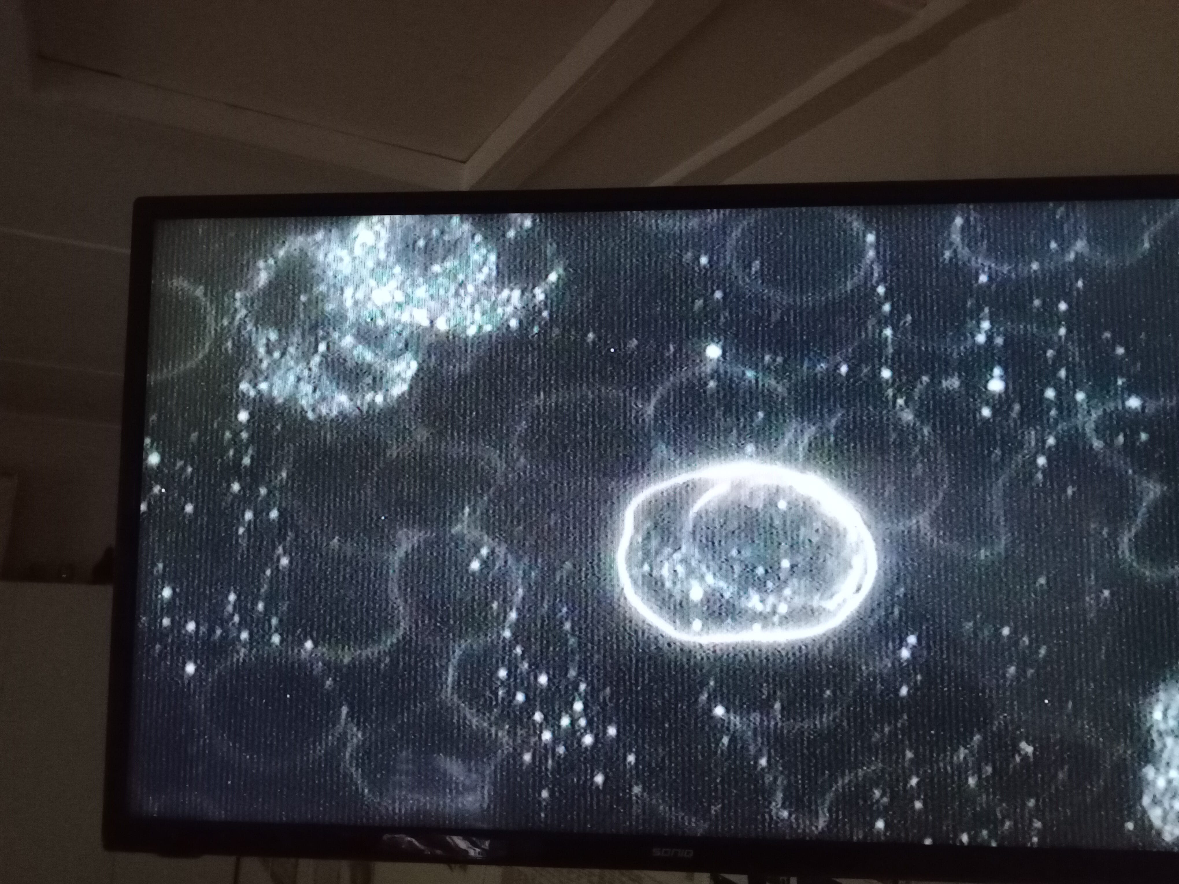
This one above appears to show the lines but it may be just the way the light is reflecting, getting harder to believe in co incidence though.
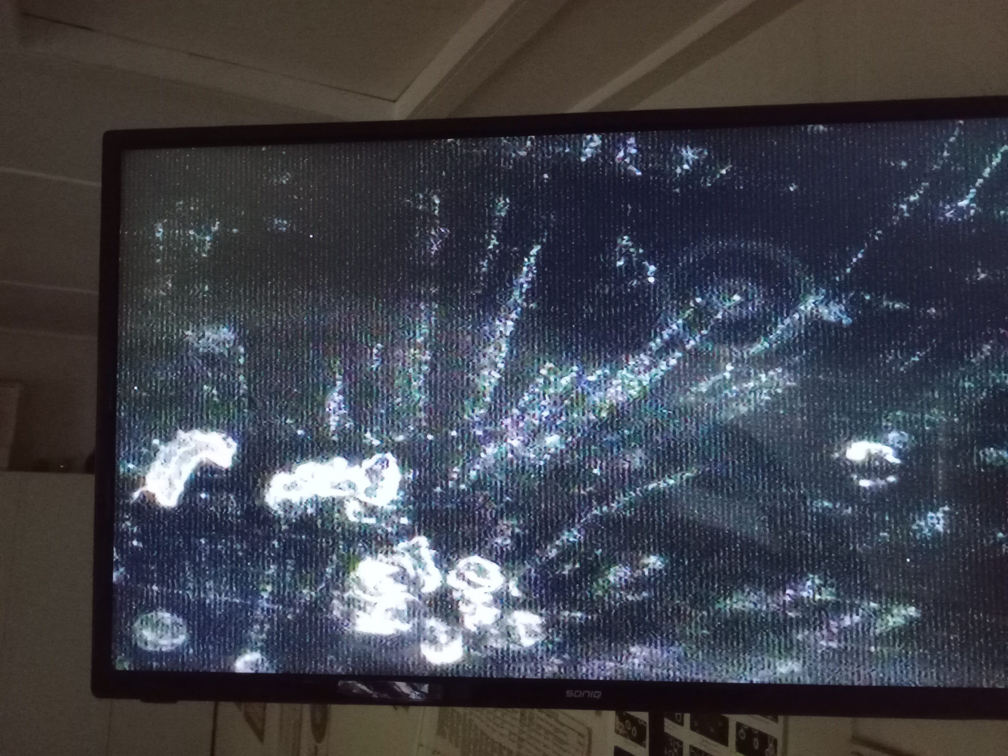
Below it may be a blood worm but idk, added both for full disclosure.
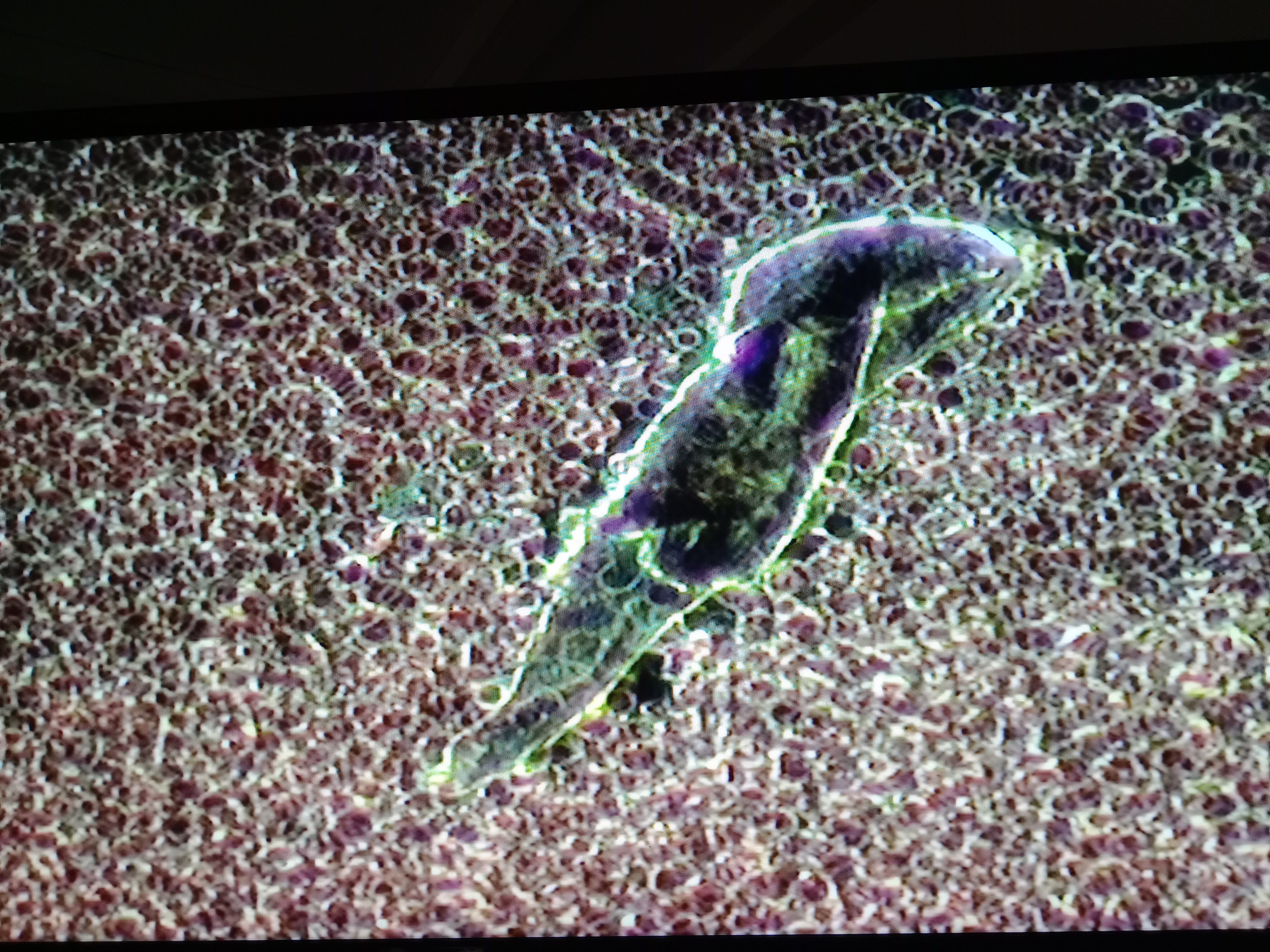
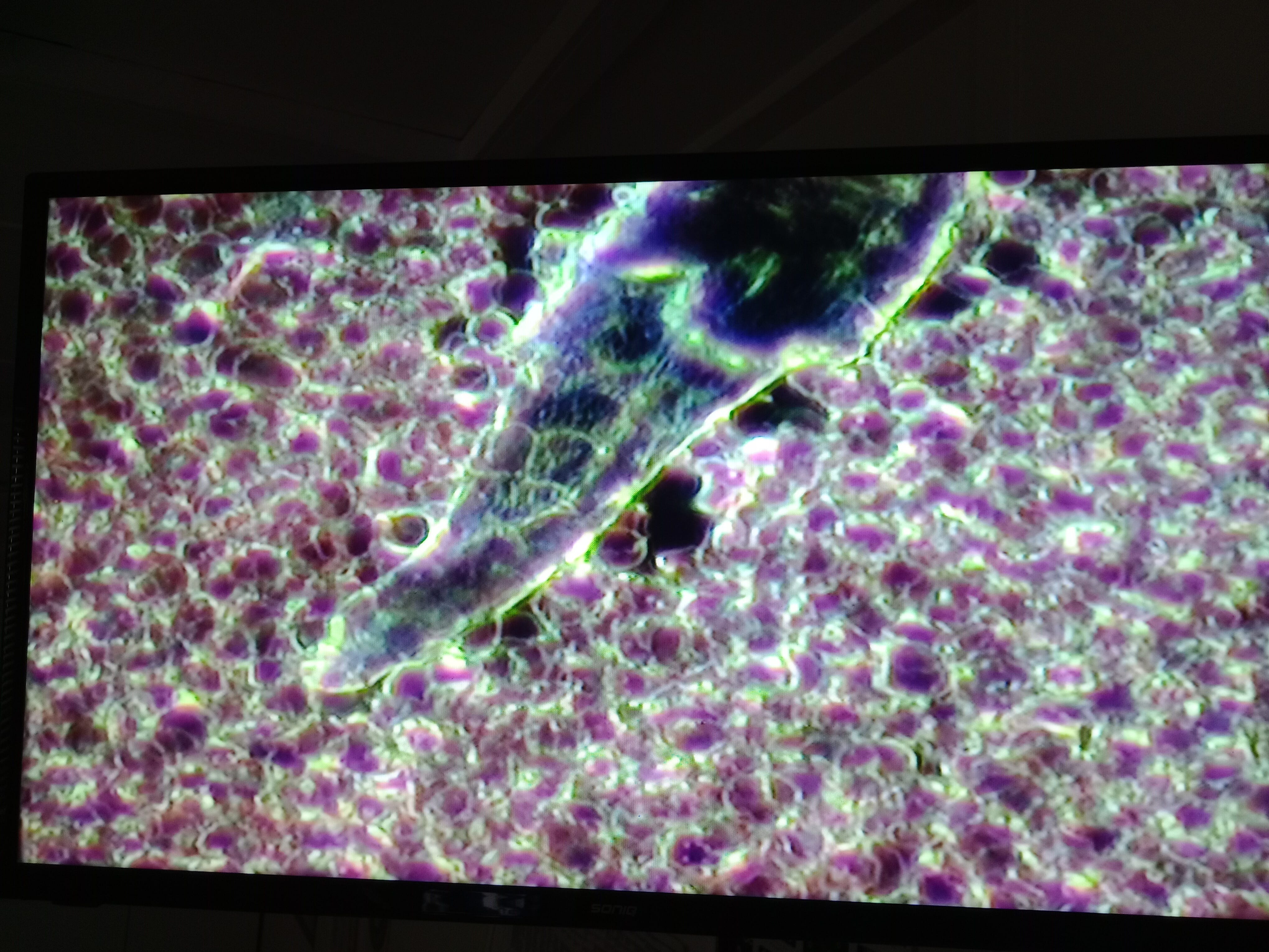
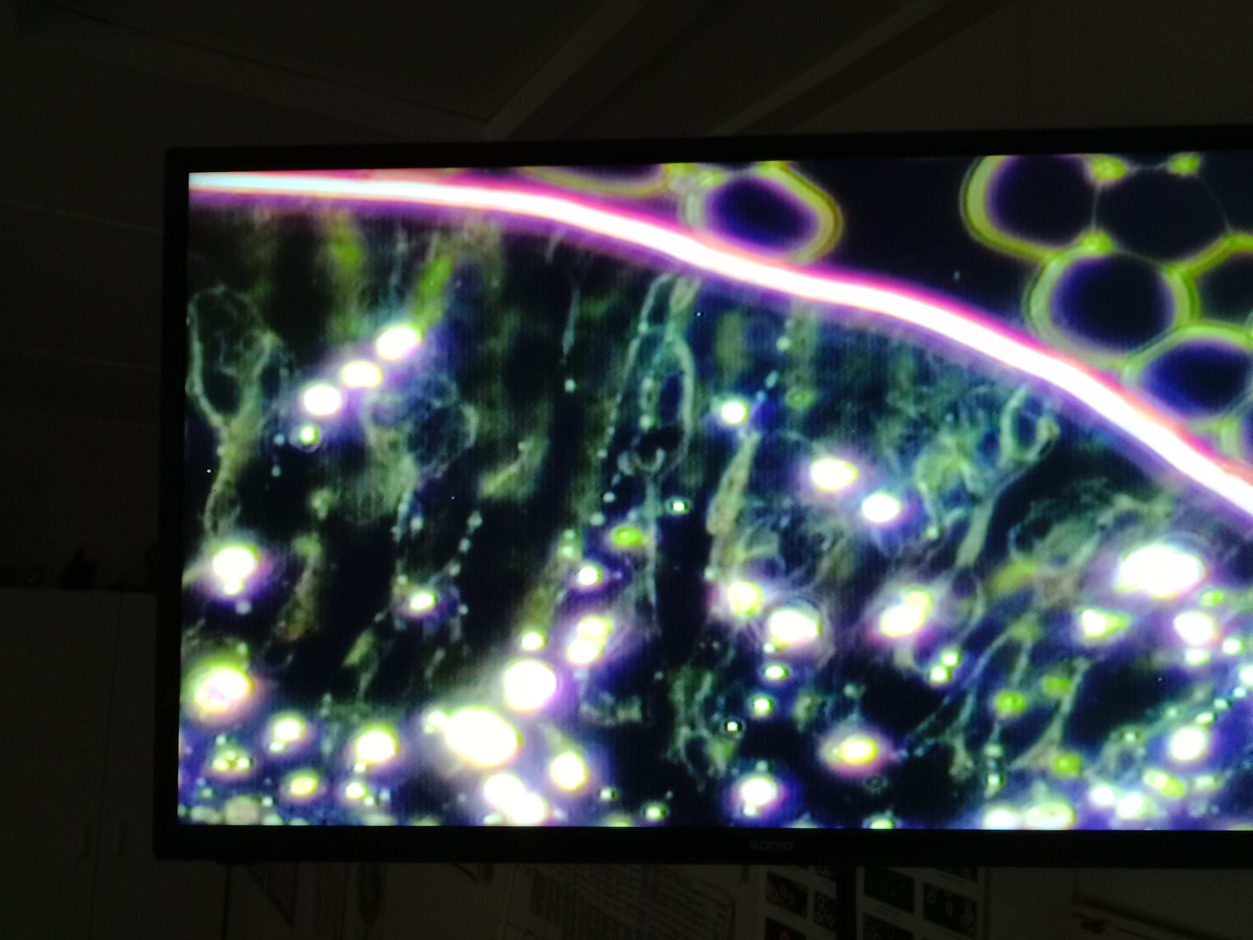
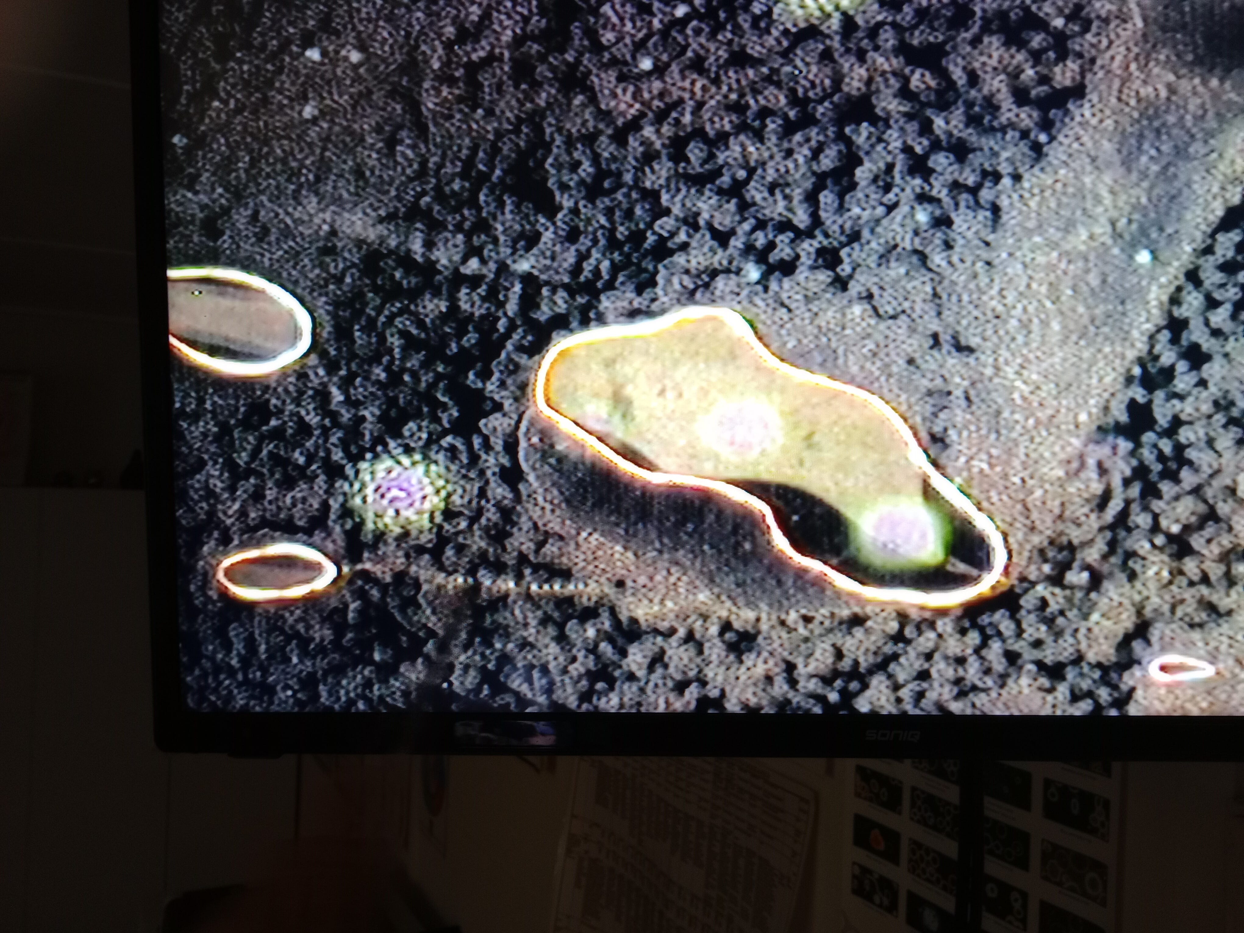
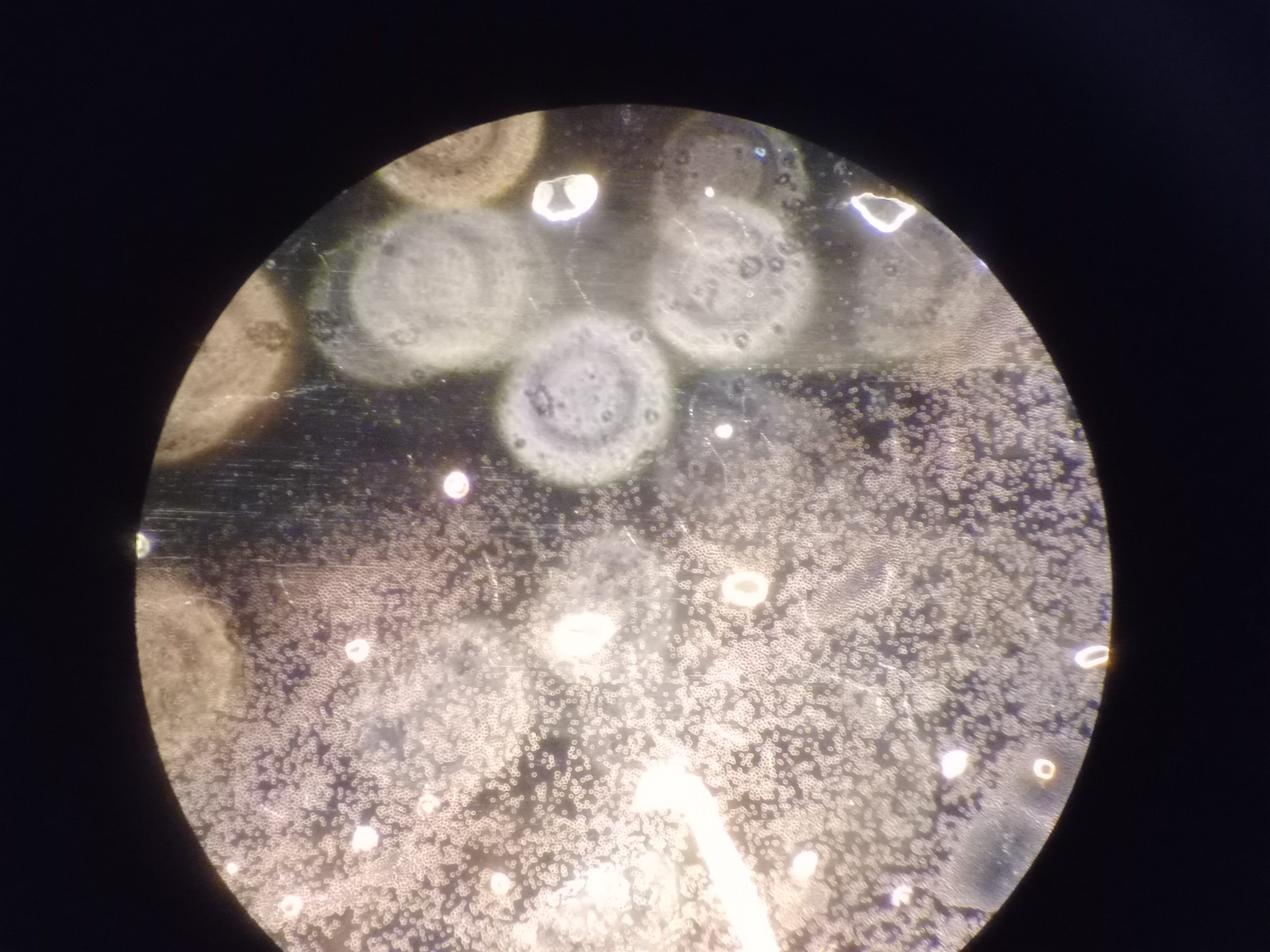
And here we have more rbc's that aren't reflecting light, my analyst calls them ghost cells which is as good as name as any. Can't see the nano tech looking thing stabbing them but that doesn't mean much.
On top of the ghost cells we have what appears to be destroyed immune cells.
In regards to the immune cell destruction? similar can be seen with the spray I showed above.
i.e.

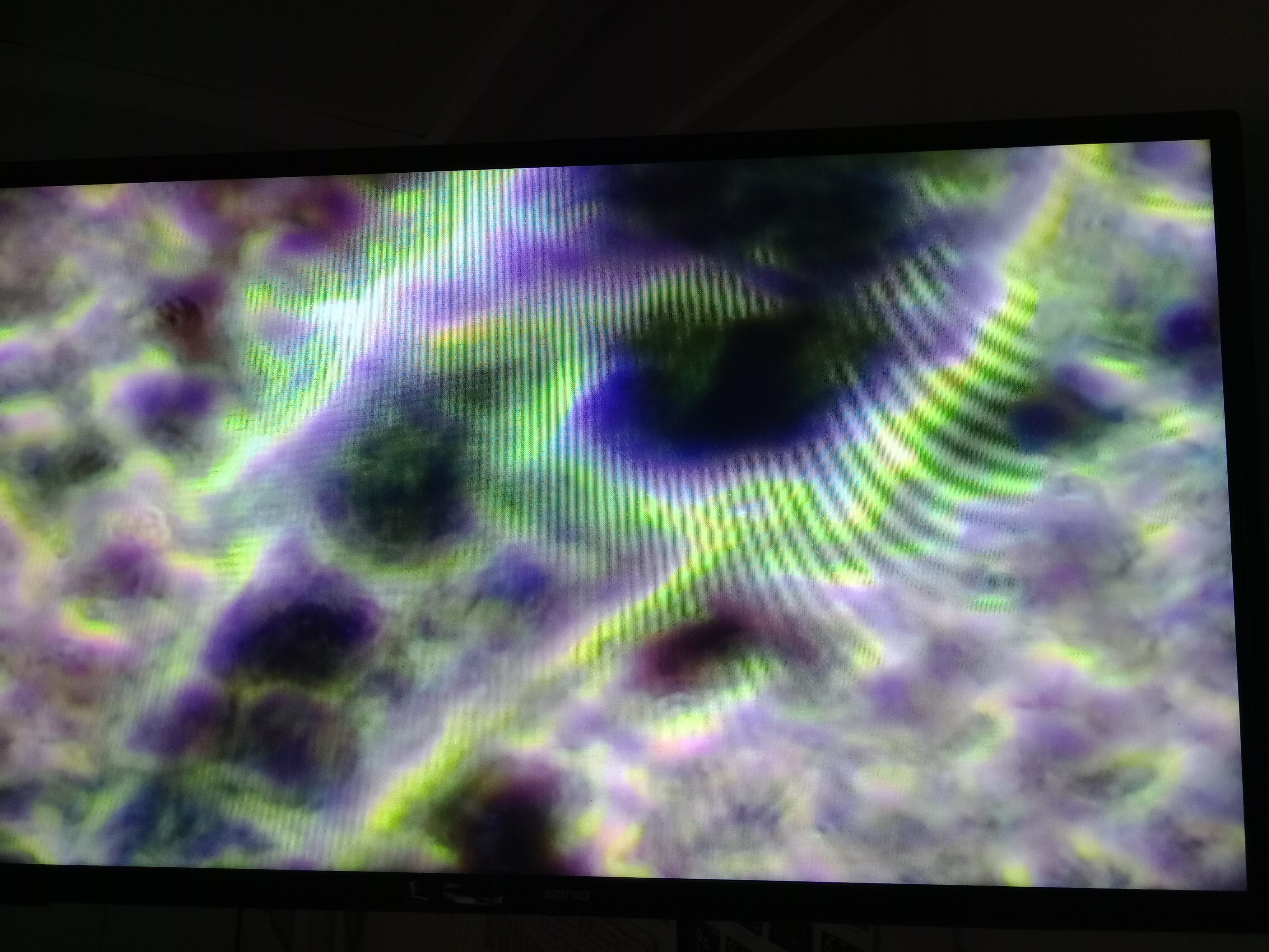
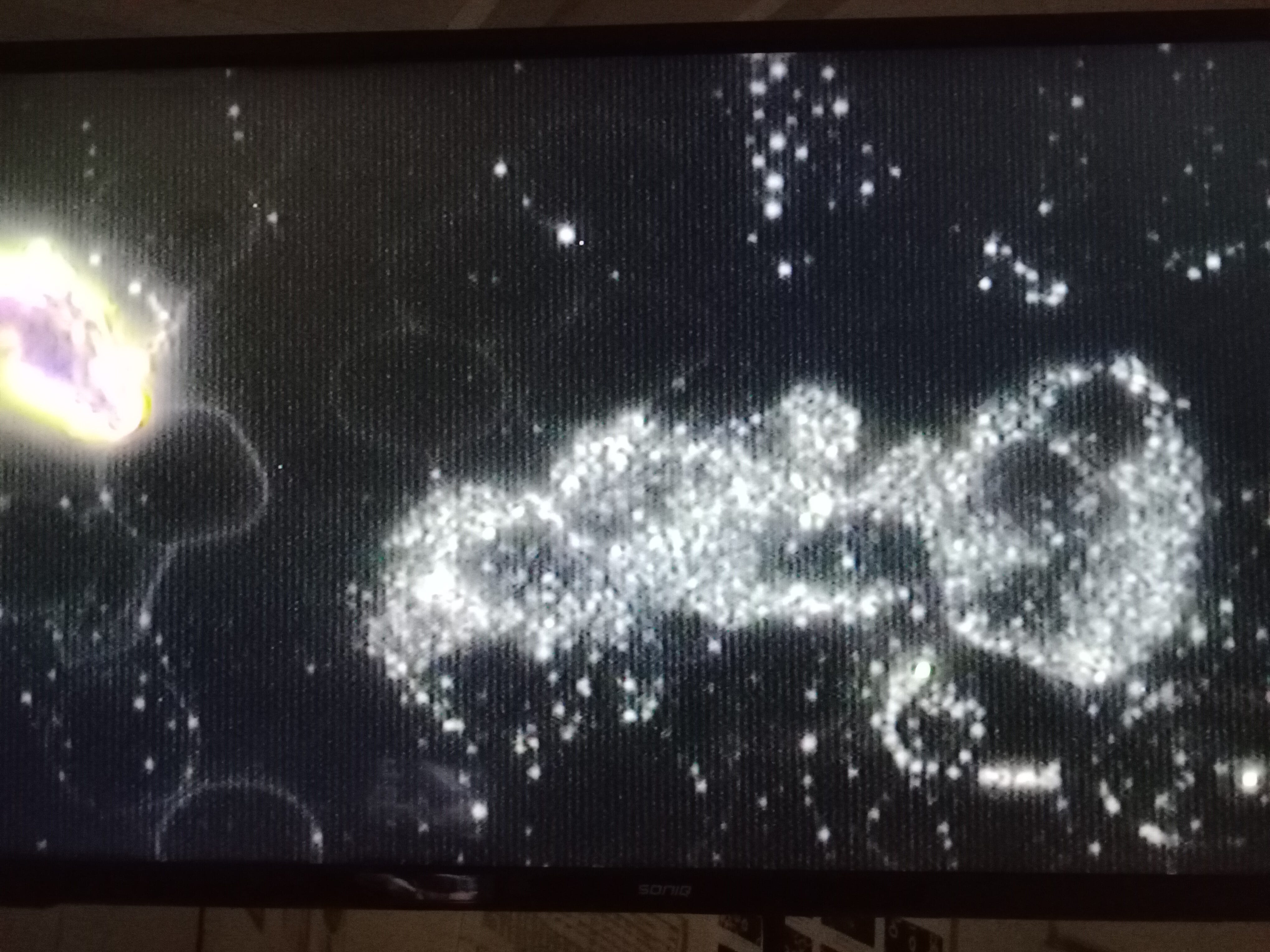
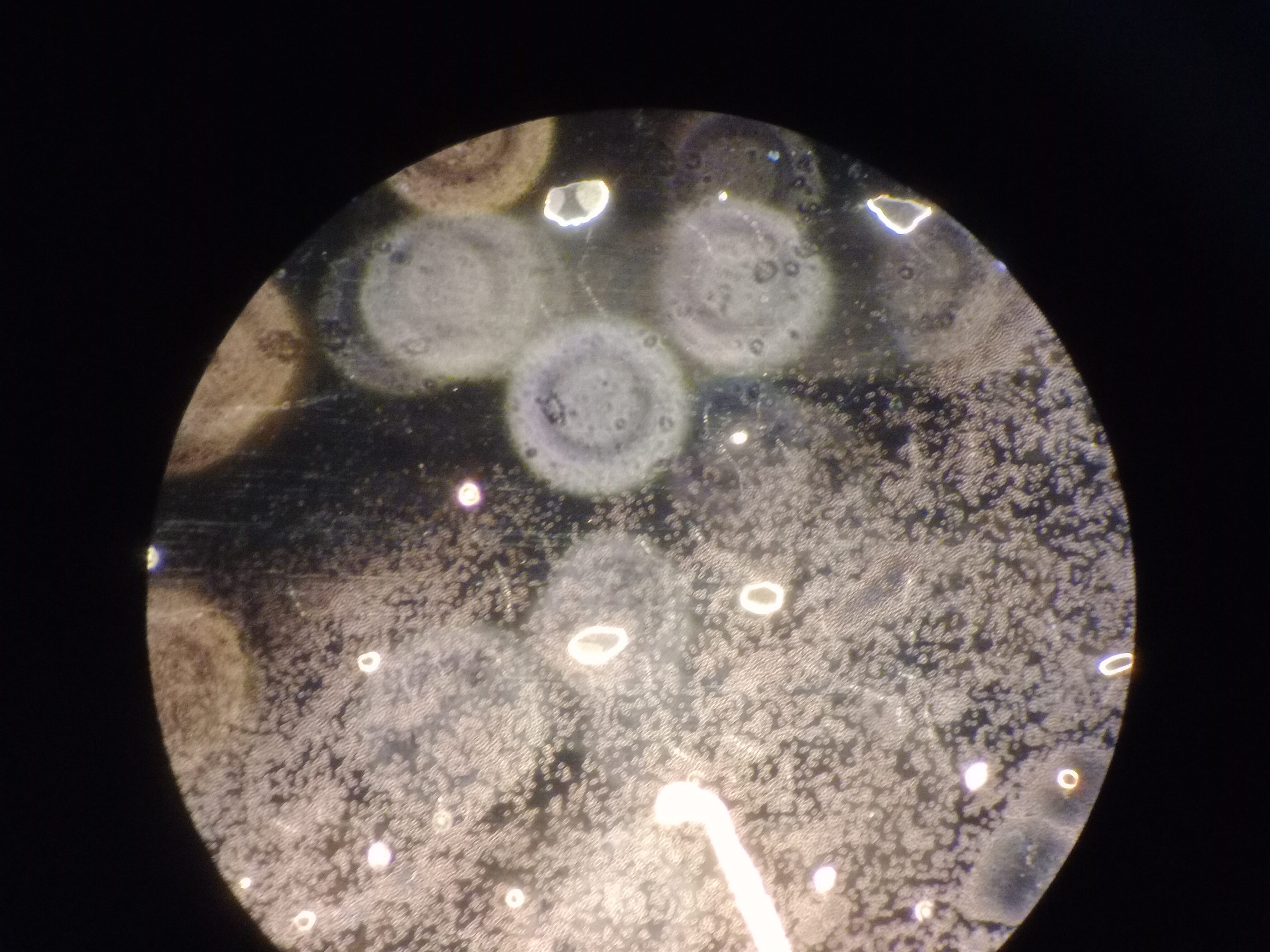
Between the microscope and the monitor there must be a colour filter to remove the yellow. Below is a photo through the eyepieces, as best I could anyway.
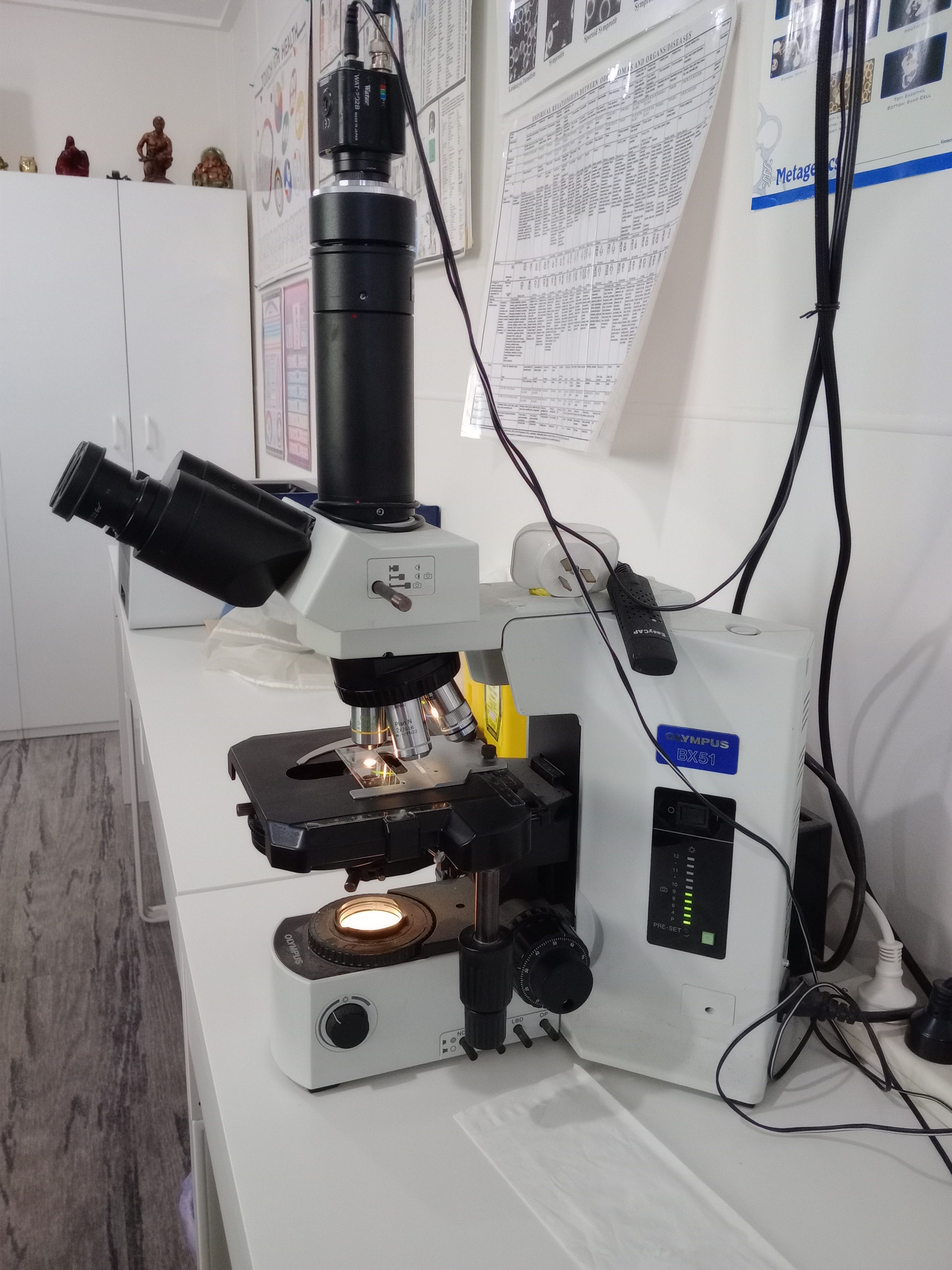
As for the microscope, of course i stuffed it up and stuck a photo at the bloody top.
Randoms


Have you seen this? https://www.2ndsmartestguyintheworld.com/p/computerized-thermographic-imaging
What I would like to know is why is it so difficult to find live blood analysis ( dark field) in my state? Also, what is the consensus on how to clear the blood?